Jet endotracheal device and its use in intubation

a technology of endotracheal device and intubation tube, which is applied in the direction of medical devices, other medical devices, respiratory apparatus, etc., can solve the problems of severe damage, difficult or impossible to intubate the trachea with grade iii or iv view quickly, and medical complications or even death

Inactive Publication Date: 2007-09-25
WEI HUAFENG
View PDF19 Cites 46 Cited by
  • Summary
  • Abstract
  • Description
  • Claims
  • Application Information

AI Technical Summary

Benefits of technology

[0022]The present invention provides an intubation device and technique for endotracheal intubation aimed at reducing the chance of hypoxia and hypercapnia and subsequent morbidity and mortality during endotracheal intubation. The intubation device and method will be especially helpful for emergency endotracheal intubation and for patients with difficult airways, such as in the cases of patients with trauma or pregnant patients who need immediate C-sections under general anesthesia.
[0040]In another aspect, the present invention provides for the use of the second or jet tube or channel for removing secretions from the trachea by the application of suction and / or for the administration of medicine, without significantly affecting ventilation through the first tube of the endotracheal unit. In yet another aspect the present invention provides for use of the second tube to monitor PetCO2 and / or airway pressure when the first tube is used for jet ventilation after endotracheal intubation has been accomplished.

Problems solved by technology

Failure to properly position an ET, or failure to insert an ET quickly, can result in severe medical complication or even death.
Failure to perform a rapid intubation after induction of general anesthesia may lead to severe damage.
It is usually easy and possible to intubate a trachea with Grade I or II view, but it is often difficult or impossible to intubate trachea with Grade III or IV view rapidly when employing only direct laryngoscopy to assist in the intubation.
It is almost impossible to perform CMV before tracheal intubation in the situation described above, because it requires a close breathing circuit for positive pressure ventilation.
However, such fiber-optic devices are usually used when the patient is awake, and not under general anesthesia.
As currently practiced, fiber-optic assisted intubation does not provide sufficient oxygenation and / or ventilation before intubation.
On the other hand, it is often difficult to intubate quickly enough a patient who is not breathing spontaneously using fiber-optic guided intubation to avoid worsening the patent's oxygenation, even at the hands of experienced medical personnel.
Other shortcomings of fiber-optic assisted intubation include the fact that the glass fibers have a tendency to break after use, and the fiber optic lens can become dislocated and are expensive to replace.
In addition, quite often secretions and blood in the airway can totally obscure the visual field.
Therefore, in the situation of an unexpectedly difficult airway, it is impractical to do an emergency fiber-optic guided intubation.
However, LMA can not be used for long-term positive pressure ventilation or to protect the airway from aspiration.
These drawbacks will limit its use in many emergency situations, such as during cardiopulmonary resuscitation, in a patient with a full stomach, and in acute respiratory failure, etc.
This requires a percutaneous puncture of large bore (>G14) catheter into trachea and is quite invasive to patient.
Often, anesthesiologists are not well trained to use transtracheal jet ventilation because of lack of practice in non-emergency situations where the method is infrequently employed because of its invasiveness and possible complications.
Meanwhile, the lack of expiratory pathway for the jet pulse in the obstructed upper airway may increase the likelihood of barotraumas.
However, the insufflation catheter is not designed as a guide for intubation, and the distal end of insufflation catheter is well positioned as a guide into trachea.
Although this arrangement permits selective ventilation of the lungs by gas currents adapted to the existing resistance conditions, it is not suitable to be utilized as a guiding catheter for intubation.
The shortcoming of this design is that the intubation guide has a fixed angle on the distal end, thus it is unable to be adapted to the different view of glottis efficiently and quickly (e.g. a throat of Grade III may be higher than in Grade II).
This makes it difficult to view the glottis with the fiber-optic device quickly in emergency situation.
In addition, it will be difficult for the distal end of oxygen supplying catheter directly facing the vocal cord opening to provide adequate oxygenation and ventilation, while at the same time to adjust the distal end of the fiber-optic device for a good view of the glottis.
No methods of oxygenation and ventilation were designed for this apparatus in case of a patient's apnea, and thus the device is impractical for use in emergency situations.
Thus, it would be difficult to use this “insufflation lumen” for generating powerful forwarding jet pulses for efficient oxygenation and ventilation if patients have apnea after receiving muscle relaxants.
The device appears to be useful for cleaning airway secretion during mechanical ventilation without interruption of oxygenation and ventilation, but does not appear to be adequate for assisting intubation in paralyzing patients with difficult airway.

Method used


Image

  • Jet endotracheal device and its use in intubation
  • Jet endotracheal device and its use in intubation
  • Jet endotracheal device and its use in intubation

Examples


Embodiment Construction

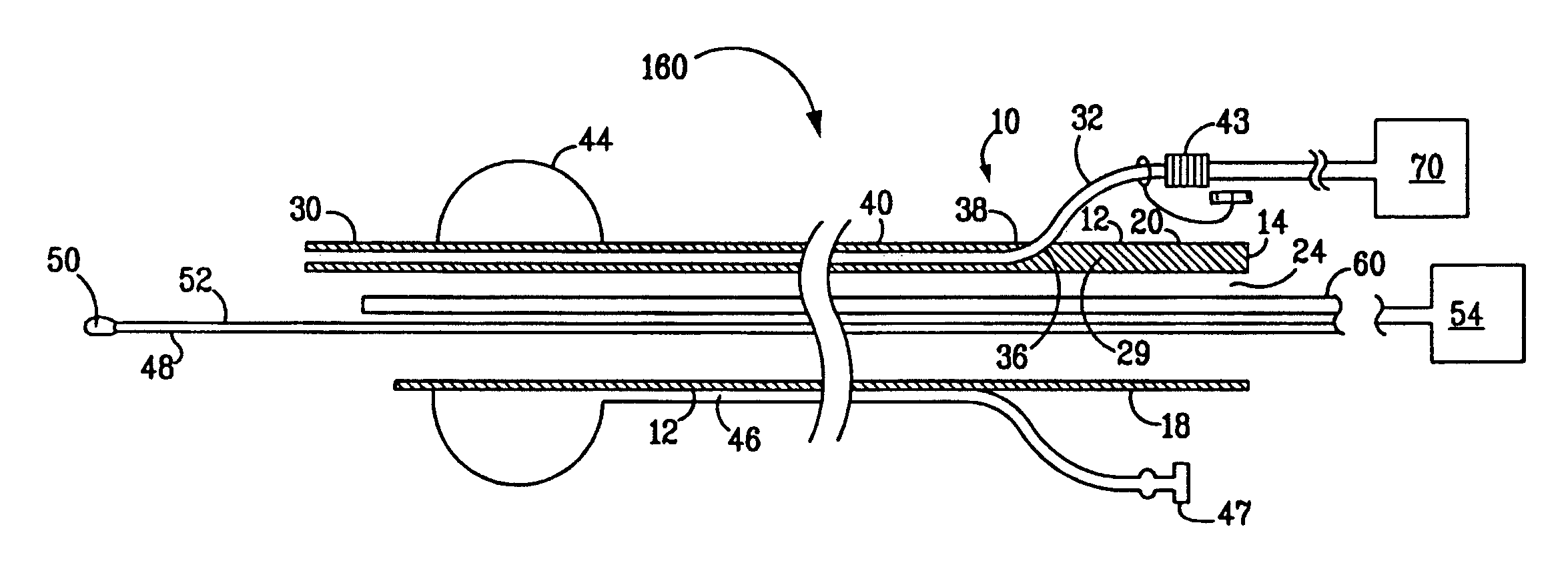
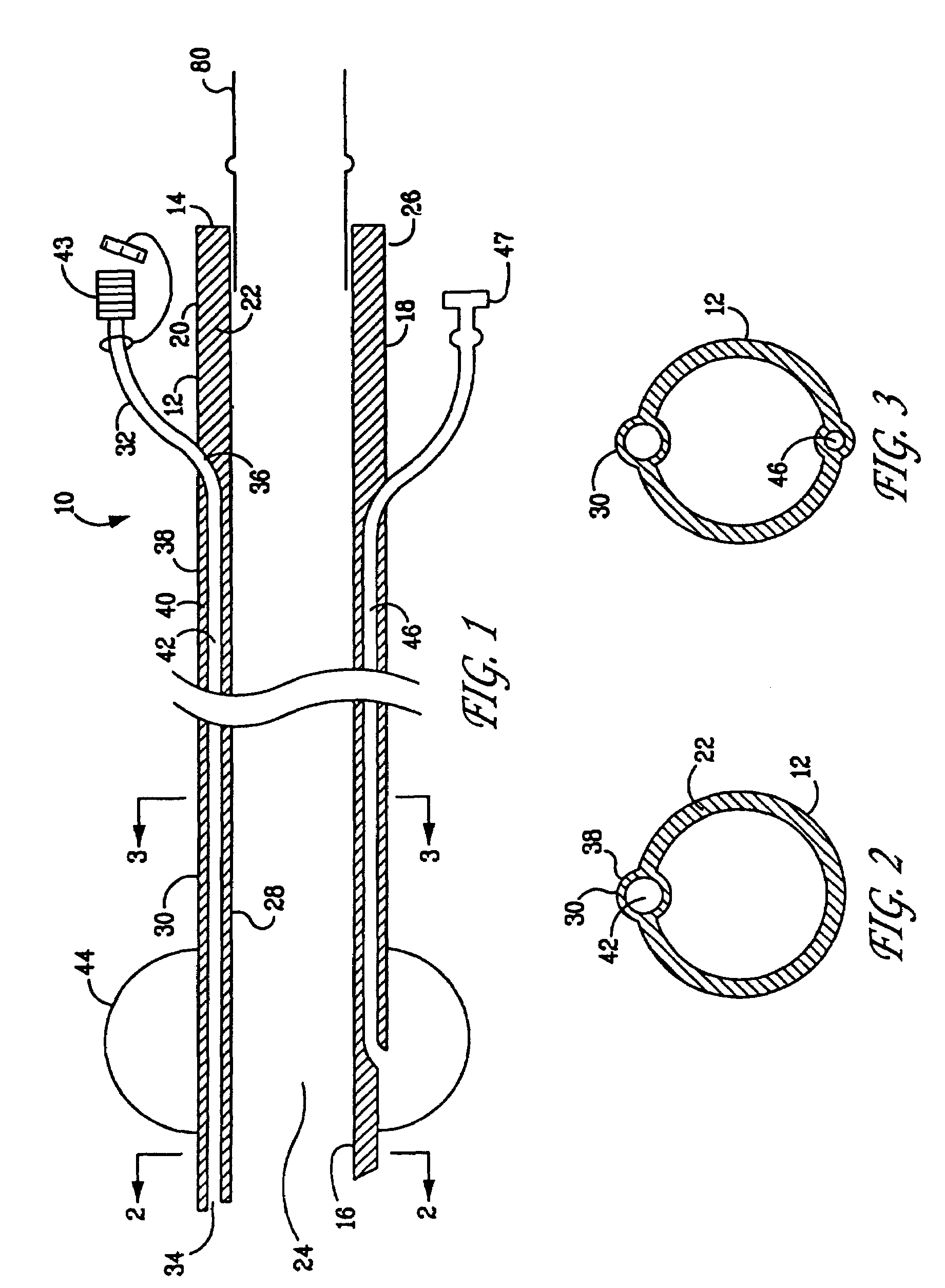
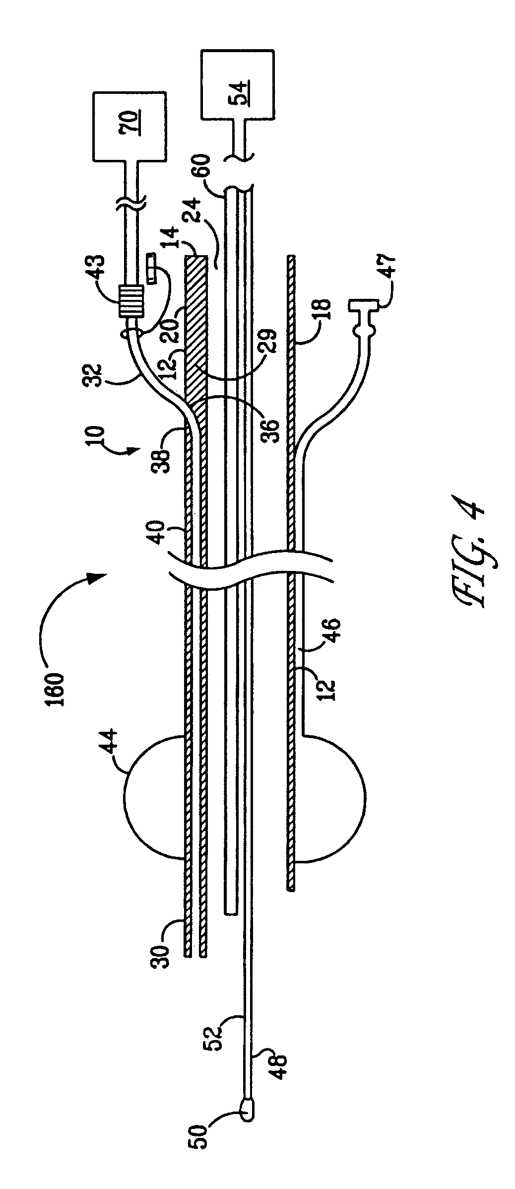
[0049]Based on experimental results obtained and described below, the endotracheal unit, device and method of the present invention provide the following advantages: The endotracheal unit and device of the present invention is simple and easy to use, even for the beginner without previous experience of tracheal intubation. Further, the endotracheal unit is not expensive and easily constructed using conventional materials. The endotracheal unit and device of the present invention provide adequate oxygenation and ventilation during the intubation even for patients with complete apnea, thus increase the safety of intubation. The endotracheal unit and device of the present invention provide easy and effective guiding methods for assisting the tracheal intubation blindly even at Grade III or IV view of glottis in a difficult airway. The endotracheal unit has been designed to minimize the intubation-associated complications, such as airway trauma, barotraumas and misplacement into the eso...


PUM


Abstract

An endotracheal device includes a unit with a pair of tubes and a beveled distal end. One tube is used for jet ventilation, while a monitoring catheter is provided inside the second, larger tube. The device is useful for otherwise difficult intubations.

Description

CROSS-REFERENCE TO RELATED APPLICATION[0001]This application claims the benefit of U.S. Provisional Patent Application No. 60 / 567,682 filed May 3, 2004.BACKGROUND OF THE INVENTION[0002]1. Field of the Invention[0003]The present invention relates to endotracheal devices and the use of such devices in the intubation of a patient.[0004]2. Brief Description of the Prior Art[0005]During surgery and other medical procedures it is often necessary to insert an endotracheal tube (“ET”) into the trachea of a patient for the purposes of ventilation assistance, or otherwise. It is sometimes necessary, such as in emergency situations of cardiac arrest, acute respiratory failure or airway obstruction etc, to position an ET rapidly. Failure to properly position an ET, or failure to insert an ET quickly, can result in severe medical complication or even death.[0006]For example, in an emergency cesarean section occasioned by fetus distress or other reasons, emergency placement of an ET in a mother's...

Claims


Application Information

Patent Timeline
Patent Type & Authority Patents(United States)
IPC IPC(8): A61M16/00A62B7/00A61M16/04A62B9/06
CPCA61M16/04A61M16/0096A61M16/0434A61M2016/0413A61M2230/432A61M16/0418A61M16/0486A61M16/0484
Inventor WEI, HUAFENG
Owner WEI HUAFENG
Why Eureka
Try Eureka