Method of lung lobe segmentation and computer system

a technology of lung lobes and computer systems, applied in the field of lung lobe segmentation, can solve the problems of not providing lobar quantitation support, and achieve the effect of accurately capturing structural features

Active Publication Date: 2008-01-01
MEVIS
View PDF14 Cites 56 Cited by
  • Summary
  • Abstract
  • Description
  • Claims
  • Application Information

AI Technical Summary

Benefits of technology

[0022]Next a lobe segmentation step is performed on the basis of the distance map. For this purpose any prior art segmentation method can be used for identification of the lobes. Application of prior art segmentation methods on the distance map rather than on the original image data provides more robust results.
[0024]In accordance with a preferred embodiment of the invention the cost function is the sum of all density values of the original image data on a path between two voxels. Preferably the end voxel of the path, which does not belong to the tubular structure, is not included in the integration of the density values of the voxels belonging to the path. This is advantageous for increased precision of the following segmentation as voxels being located on the lobar fissures do have a slightly higher density value in comparison to their neighboring voxels. Excluding the voxel at the end point of the path therefore improves the performance of the cost function.
[0027]In accordance with a further preferred embodiment of the invention the world coordinates of the voxels are used for calculating the distance measure. This enables to more accurately capture the structural features which are represented by the image data. For compensation of the anisotropic voxels resulting from certain image data acquisition techniques the image data is remapped to the world coordinate system prior to calculating the distance measure.
[0028]In accordance with a further preferred embodiment of the invention a weighted combination of the distance map and the original image data is performed. This has the advantage that the sharp fissure contours of the original image data are preserved in the combined image.

Problems solved by technology

Such prior art lung segmentation techniques do not take the the lung lobes into account and thus do not provide lobar quantitation support.

Method used


Image

  • Method of lung lobe segmentation and computer system
  • Method of lung lobe segmentation and computer system
  • Method of lung lobe segmentation and computer system

Examples


Embodiment Construction

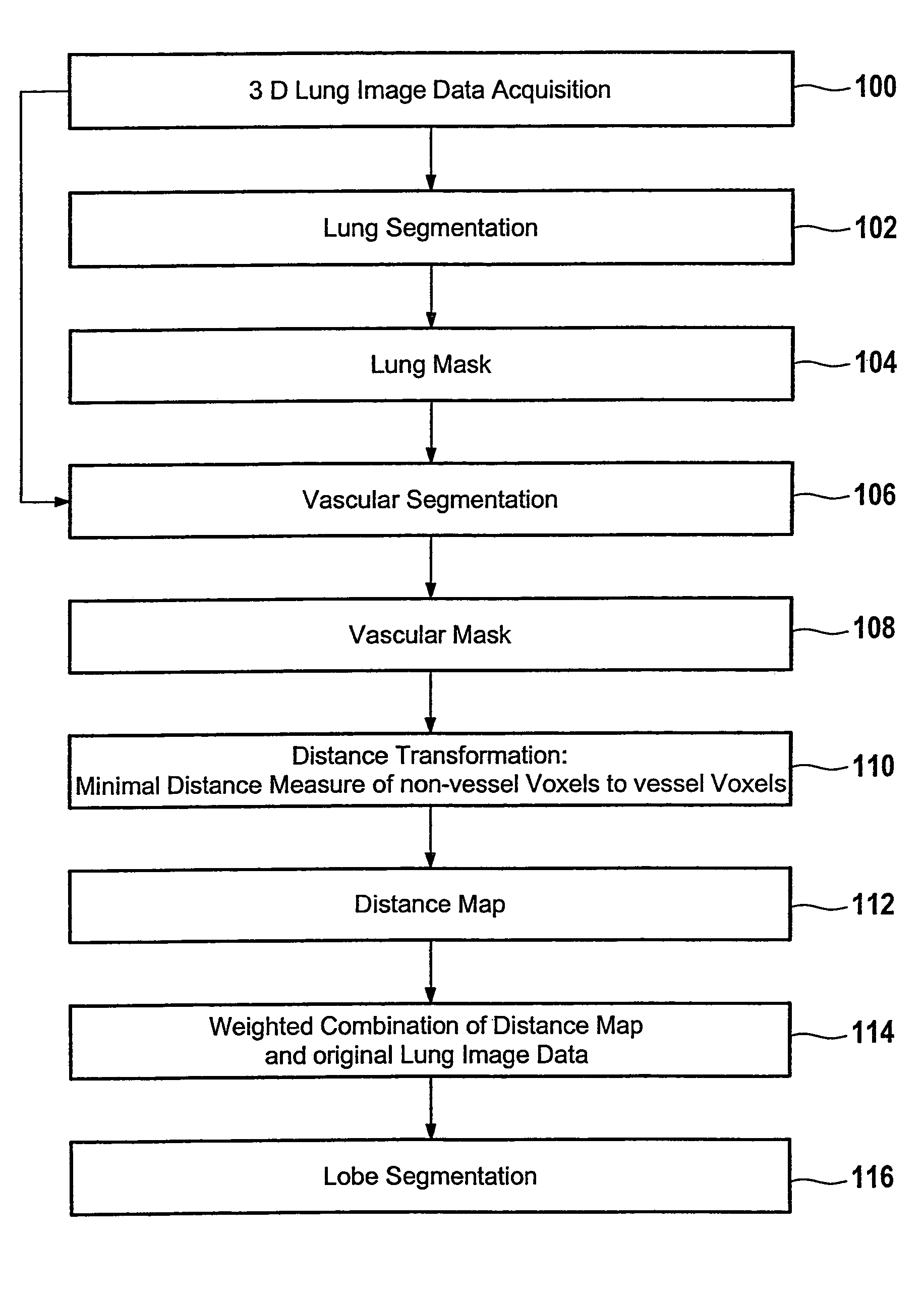
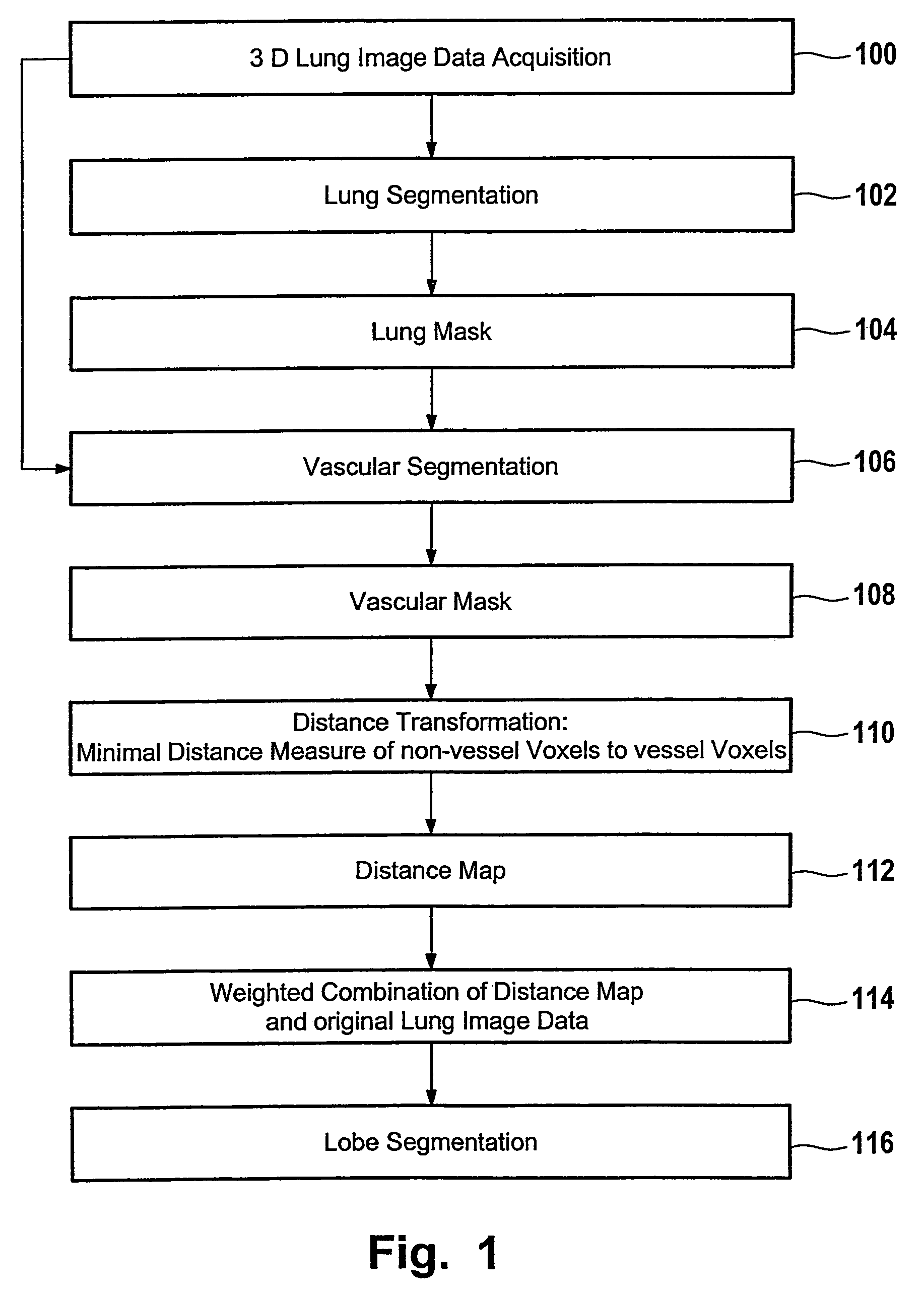
[0040]FIG. 1 shows a flow chart for lung lobe segmentation. In step 100 three dimensional lung image data is acquired by computer tomography, nuclear magnetic resonance tomography or by means of another image modality. For example thoracic image data is acquired from chest computer tomography data.

[0041]Without restriction of generality it is assumed that the image data covers an input volume V of dimensions X×Y×Z. The set of all voxel positions within the input volume is defined as V:={0, . . . , X−1}×{0, . . . , Y−1}×{0, . . . , Z−1} whilst I:=(iv)v∈V refers to the 3D input matrix. An entry iv denotes the integer density value of the voxel v within the input volume. For example, values range from 0 to 4095 corresponding to Hounsfield Units (HU) between −1024 HU and 3071 HU. In case the world coordinates of voxel v are used this in indicated by world (v).

[0042]In step 102 a lung segmentation procedure is performed on the image data I, if necessary. Performing such a lung segmentati...


PUM


Abstract

A method of lung lobe segmentation having the steps of: providing of three dimensional first image data of a lung; segmentation of a tubular structure to identify first voxels of the first image data belonging to the tubular structure and to identify second voxels of the first image data which do not belong to the tubular structure; determining a distance measure for each one of the second voxels, by determining a minimum distance between each one of the second voxels to one of the first voxels to provide second image data; and performing a lobe segmentation based on the second image data.

Description

BACKGROUND OF THE INVENTION[0001]1. Field of the Invention[0002]The present invention relates to the field of segmentation of medical digital images, and more particularly to lung lobe segmentation.[0003]2. Prior Art[0004]The human lungs are composed of distinct anatomic compartments called lobes. Since they represent functional units, the lobes have their own bronchial and vascular systems. These systems are solely connected close to the hilus, which is defined as the area where bronchi as well as the pulmonary arteries and veins enter the lungs. The lobes in each lung are separated by thin tissue, the so-called lobar fissures. An oblique fissure and a horizontal fissure divide the right lung into upper, lower, and middle lobe. In the left hand lung there is only an oblique fissure separating lower and upper lobe.[0005]High-resolution X-ray computer tomography (CT) is the standard for pulmonary imaging. Depending on the scanner hardware, CT can provide high spatial and high tempora...

Claims


Application Information

Patent Timeline
Patent Type & Authority Patents(United States)
IPC IPC(8): G06T17/40G06V10/26
CPCG06K9/34G06K2209/051G06V10/26G06V2201/031
Inventor KUHNIGK, JAN-MARTIN
Owner MEVIS
Why Eureka
Try Eureka