Methods and systems to detect and classify retinal structures in interferometric imaging data

a technology of interferometric imaging and classification methods, applied in image analysis, image enhancement, instruments, etc., can solve the problems of inconvenient or even impossible manual segmentation of human operators in high-throughput clinical environments, high computational and execution time requirements, and high difficulty in segmentation, so as to reduce the number of redundant processing and reduce the segmentation error

Active Publication Date: 2019-04-11
CARL ZEISS MEDITEC INC
View PDF4 Cites 2 Cited by
  • Summary
  • Abstract
  • Description
  • Claims
  • Application Information

AI Technical Summary

Benefits of technology

[0009]Systems and methods of the present application are directed towards improved segmentation of interferometric image data. In one aspect of the present invention, a workflow in which OCT data is preprocessed to be divided into regions of common size, resolution, alignment, intensity and statistical properties is described. Such division allows the processing of image data of different extent, scan pattern acquisition parameters, complexity, illumination conditions and pathologies by the same segmentation workflow in a reliable and controlled manner. Processing divided regions of such characteristics independently provides more robust results and decreases the computational requirements of the segmentation algorithm. In addition, constraining each segmentation problem to regions of data having reduced span and common characteristics allows for the statistical properties of the pixels surrounding a particular retinal layer to be more similar within the region, which helps in the association of locations belonging to the same retinal layer.
[0013]The various aspects / embodiments of the present application discussed above are able to locate and to associate features with different retinal structures or layers based upon characteristic information: location, connectedness, and statistical properties. This includes the possible variation of boundaries of anatomical structures as well as textural information of those structures. The preprocessing step in which the data is divided into regions of common statistical properties that are processed independently allows each identifiable feature within a region to share common characteristic information. This approach offers greater flexibility over the fixed constraints on layer smoothness, since it learns from the data what amount of variability is possible and in what regions, while on the other hand, constrains data to a plausible space of states. Moreover, the methods and systems discussed herein are completely self-contained in learning: meaning that each image provides the information required to train a classifier. Note that a dataset can consist of a single B-scan, multiple B-scans at the approximate same location, or it can be volumetric, comprising a plurality of B-scans at multiple locations. In the case of volumetric datasets, the information derived from the features of a B-scan can be used to identify the features of an adjacent scan, in short, e.g. the statistical metrics of one B-scan could be used for subsequent B-scans in a vicinity of the B-scans.
[0016]A unified or coherent segmentation system or method needs to accommodate any retinal scan, whether the contrast be low or high, or one beset with pathological irregularities. Thus, the present application embodies a common framework for: the preprocessing and processing of images, and, the detection and classification of features to retinal structures. These embodiments are dynamic and adaptable, which significantly reduces redundant processing, while also reducing the segmentation error.

Problems solved by technology

However, due to the sheer amount of data, it is inconvenient or even impossible for a human operator to manually perform the segmentation in a high throughput clinical environment.
In general, segmentations tend to be very sensitive to noisy data or are limited only to a small number of layers.
The previously mentioned segmentation approaches are beset with one or more of the following disadvantages: 1) they distinguish only the most prominent layers, 2) do not exhibit robustness in noisy and varied cases, 3) often require manual intervention of the operator; 4) are specific to a subset of retinal layers, 5) cannot deal with the range of retinal pathologies that could be present, 6) are computationally and execution-time intense, and 7) often require a classification system based upon an external reference database.

Method used


Image

  • Methods and systems to detect and classify retinal structures in interferometric imaging data
  • Methods and systems to detect and classify retinal structures in interferometric imaging data
  • Methods and systems to detect and classify retinal structures in interferometric imaging data

Examples


Embodiment Construction

[0031]All patent and non-patent references cited within this specification are herein incorporated by reference in their entirety to the same extent as if the disclosure of each individual patent and non-patient reference was specifically and individually indicated to be incorporated by reference in its entirely.

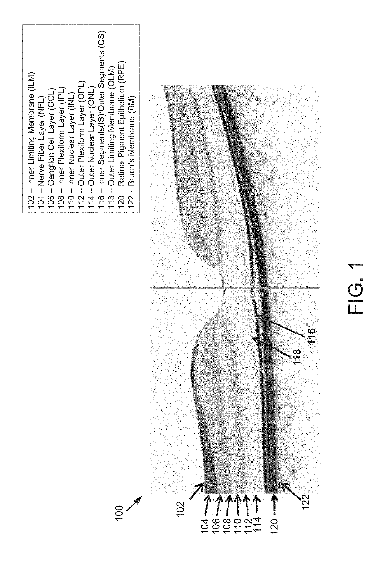
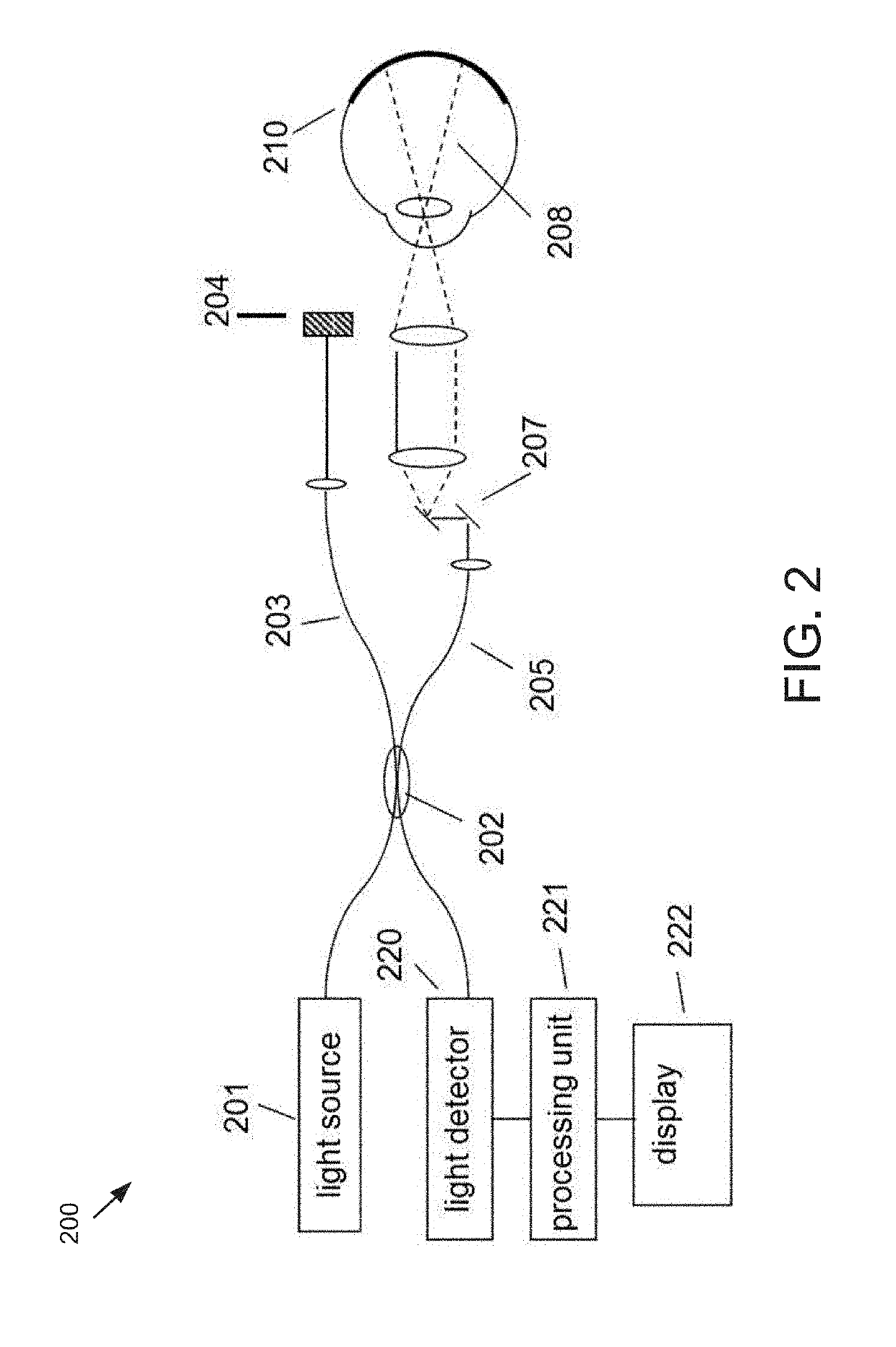
[0032]FIG. 2 illustrates a generalized Fourier Domain optical coherence tomography (FD-OCT) system 200 that is used to collect an OCT dataset suitable for use with the present set of embodiments. As depicted, the FD-OCT system 200 includes a light source 201, typical sources including, but not limited to, broadband light sources with short temporal coherence lengths or swept laser sources. Light from source 201 is routed, typically by optical fiber 205, to illuminate the sample 210, which could be any of the tissues or structures within an eye. The light is scanned, typically with a scanner 207 between the output of the fiber and the sample, so that the beam of light (dashed...


PUM


Abstract

Methods and systems are presented to analyze a retinal image of an eye and assigns features to known anatomical structures such as retinal layers. One example method includes receiving interferometric image data of an eye. A set of features is identified in the image data. A first subset of identified features is associated with known retinal structures using prior knowledge. A first set of characteristic metrics is determined of the first subset of features. A second set of characteristic metrics is determined of a second subset of features. Using the characteristic metrics of the first and the second sets, the second subset of features is associated with the retinal structures. Another example method includes dividing interferometric image data into patches. The image data in each patch is segmented to identify one or more layer boundaries. The segmentation results from each patch are stitched together into a single segmentation dataset.

Description

PRIORITY[0001]This application is a divisional application of U.S. patent application Ser. No. 15 / 246,036, filed Aug. 24, 2016, which claims priority to U.S. Provisional Application Ser. No. 62 / 210,953, filed Aug. 27, 2015, the contents of each of which is hereby incorporated by reference.TECHNOLOGICAL FIELD[0002]The present application concerns improved methods and systems for detecting and identifying structures in images obtained with interferometric imaging systems.BACKGROUND[0003]A wide variety of interferometric imaging techniques have been developed to provide high resolution structural information in a wide range of applications. Optical Coherence Tomography (OCT) is a technique for performing high-resolution cross-sectional imaging that can provide images of samples including tissue structure on the micron scale in situ and in real time. OCT is an interferometric imaging method that determines the scattering profile of a sample along the OCT beam by detecting light reflecte...

Claims


Application Information

Patent Timeline
Patent Type & Authority Applications(United States)
IPC IPC(8): G06T7/00G06V10/44G06V10/764
CPCG06T2207/30041G06T7/0012G06T2207/10101G06T2207/20081G06T2200/04G06T2207/20021G06T2207/20076G06T7/12G06T7/143G06T7/181G06V10/44G06V2201/03G06V10/761G06V10/764G06F18/22G06F18/2415
Inventor BAGHERINIA, HOMAYOUNDE SISTERNES, LUIS
Owner CARL ZEISS MEDITEC INC
Why Eureka
Try Eureka