
Sample preparation integrated chip

a sample preparation and chip technology, applied in the field of apparatus and assay systems, can solve the problems of cumbersome microarray-based assays, high manufacturing cost of chips, inefficiencies in white blood cell isolation, etc., and achieve the effect of facilitating the introduction and flow of sample fluid

Inactive Publication Date: 2008-03-04
NANYANG TECH UNIV +1
View PDF53 Cites 208 Cited by
  • Summary
  • Abstract
  • Description
  • Claims
  • Application Information

AI Technical Summary

Benefits of technology

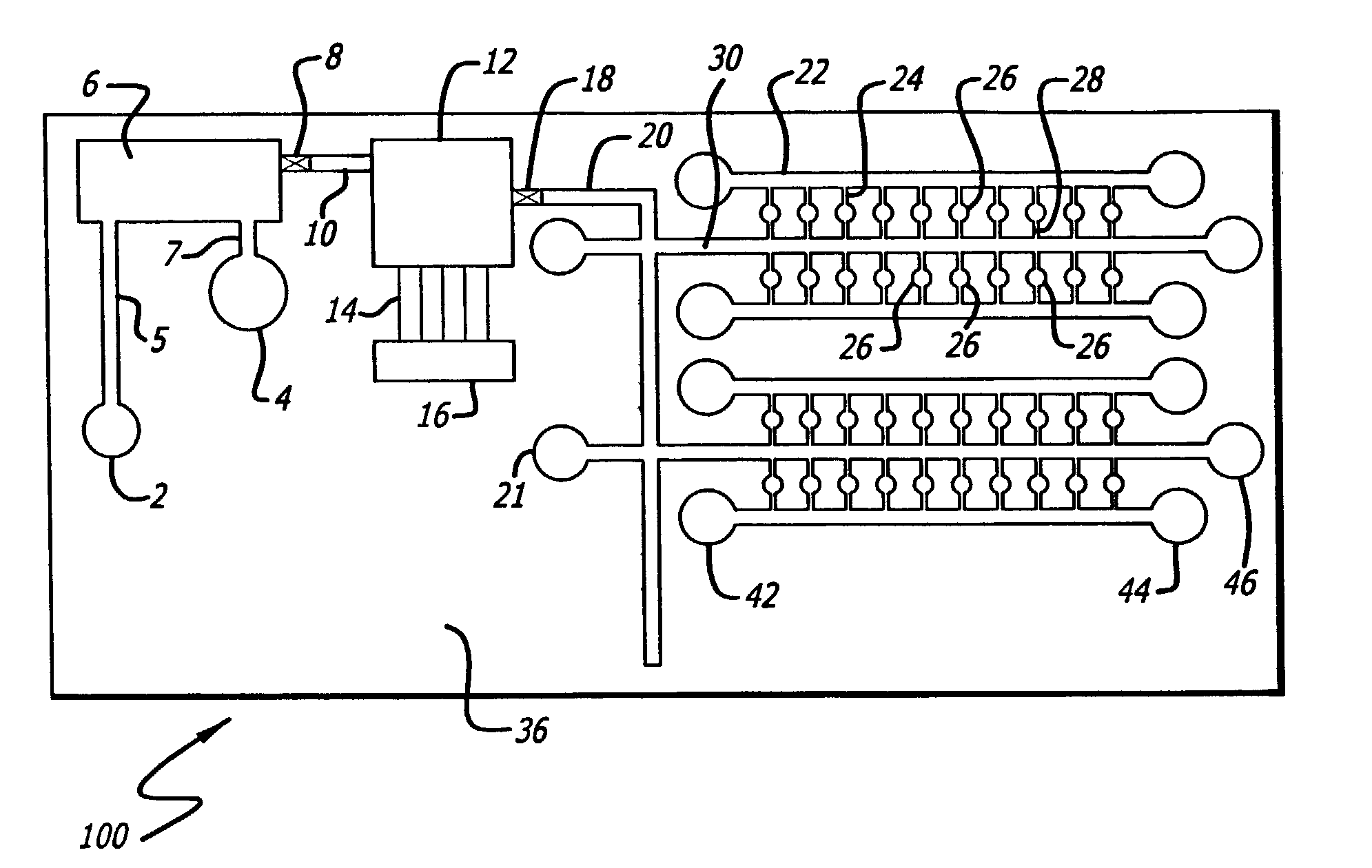
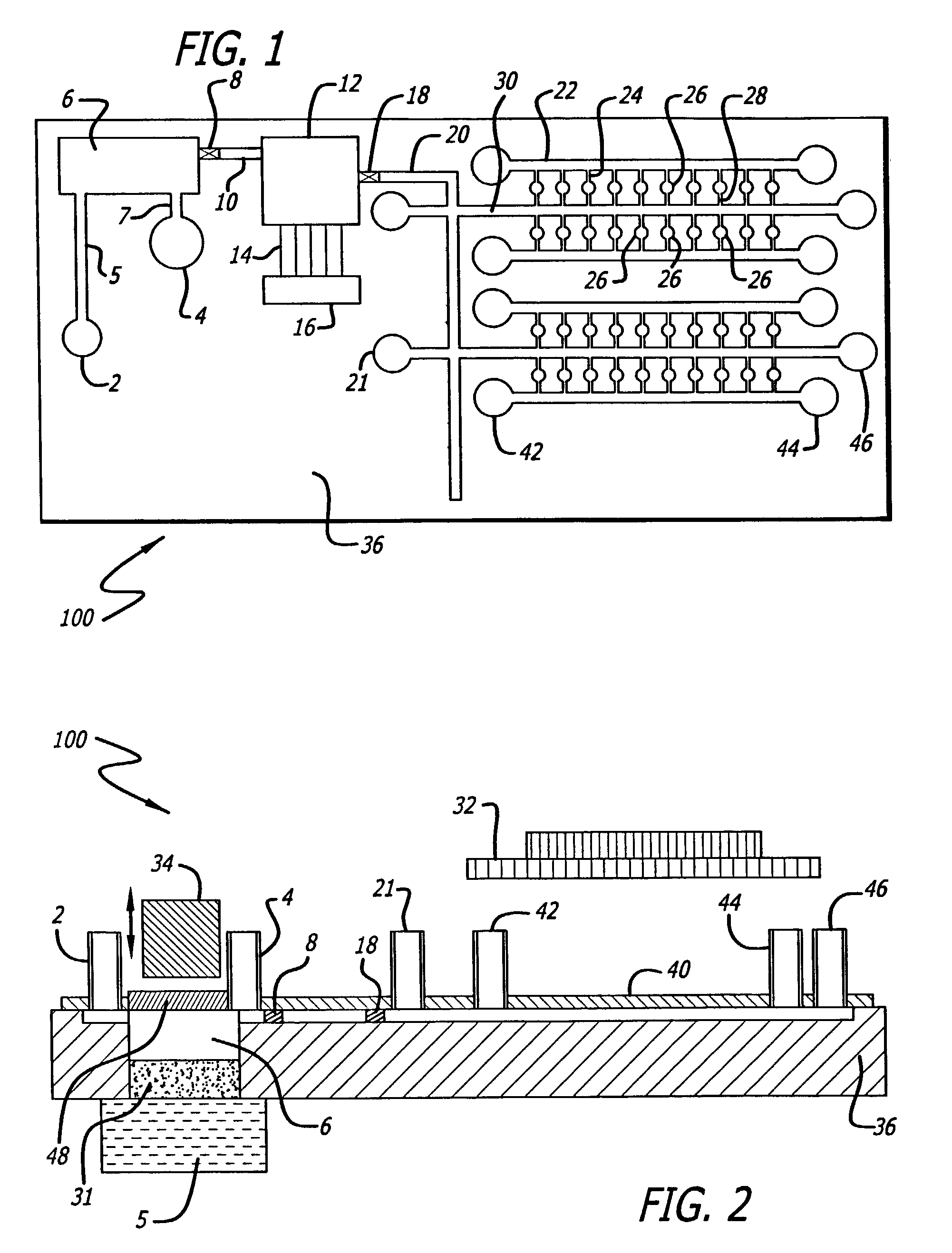
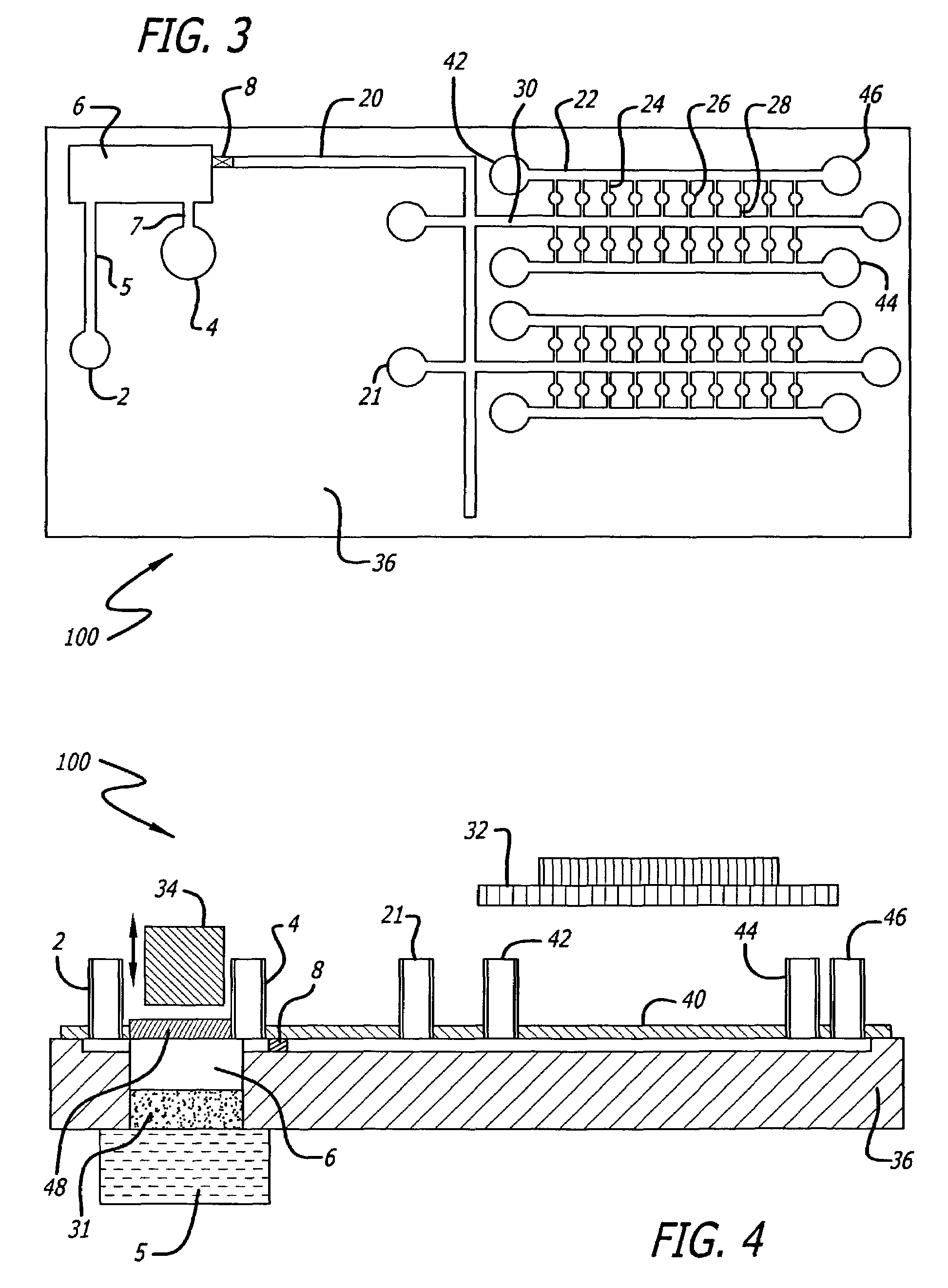
The present invention is a microchip apparatus that can detect and diagnose diseases in a subject by using various types of assays. The apparatus has a sample preparation chamber, a sample introduction inlet, a buffer introduction inlet, a flow-promoting fluid chamber, a storage chamber, and an isolation device for isolating and permitting flow of a fluid from the sample preparation chamber to the assay station. The apparatus can also include an isolation device for isolating and permitting flow of a fluid from the flow-promoting fluid chamber to the assay station. The method involves depositing a specific DNA fragment in the assay station and drying it, injecting a biological blood sample and a washing buffer into the sample preparation chamber, injecting a lysing buffer and a gas into the sample preparation chamber, diffusing a chemical from the chemical storage chamber into the flow-promoting fluid chamber, causing the isolation device to permit flow of the lysing buffer containing DNA fragments into the first multi-purpose channel to the assay station, detecting when the assay station is filled with the lysing buffer containing the DNA fragments, amplifying the DNA fragments, and detecting the amplified DNA fragments.

Problems solved by technology

Red blood cells pass through the micro-filters but tend to clog up the filters over time causing inefficiencies in white blood cell isolation.
The Yuen, et al. microchip employs a microtemperature sensor, making the Yuen, et al. chip expensive to fabricate.
The microarray-based assays are cumbersome, taking about a day to complete and requiring standalone equipment to conduct sequential batch analyses.
Rapid diagnoses are precluded and current microarray devices do not permit sample preparation to be integrated onto the chip.
Additional disadvantages of the current on-chip DNA analysis systems have recently been reported.
Such disadvantages include lack of sample injection ability, poor DNA isolation and inability to conduct multiple PCR analyses.
Current nucleic acid methods using chips typically require complex off-chip sample DNA isolation, integrated micro-heaters and micro-temperature sensors for PCR thus making current chips and associated methods of using same very expensive and non-disposable.

Method used


Image

  • Sample preparation integrated chip
  • Sample preparation integrated chip
  • Sample preparation integrated chip

Examples


example 1

[0216]Antibody-antigen fluorescence quenching assay: An antibody was labeled with OG-514 (Oregon green 514 carboxylic acid, succinimidyl esters) and the antigen (peptide, protein, whole cells, carbohydrate, aptamers, etc.) was labeled with QSY-7 (QSY-7 carboxylic acid, succinimidyl esters). Fluorescence quenching prevented or suppressed the detection of OG-514 fluorescence. The labeled antibody-antigen complex was lyophilized in the assay stations. Sample fluid preparation releases proteins or other antigens into PBS or TBS buffer with or without detergent (e.g. Tw-20 or Triton-X 100) of various concentration (e.g. 0.05% Tw-20 and 1%Triton-X-100), and flow into the assay station(s) via channels. Upon re-hydration in the assay station, the labeled antibody-antigen complex participates in competitive reaction with the unlabeled antigen. Competition with unlabeled antigen releases the OG-514 labeled antibody whose fluorescence is detected at about 528-530 nm.

example 2

Double Sandwich Antibody FRET

[0217]Two monoclonal antibodies directed against 2 non-competitive epitopes of the CD8-alpha chain were utilized. One of the monoclonals was labeled with the dye phycoerythrin (PE) and the other allophycocyanin (APC).

[0218]FRET was observed when excitation light was directed to PE but the efficiency was only 10%. Reference: Batard P., et.al., Cytometry Jun. 1, 2002;48(2):97-105. The efficiency of FRET may be improved by using near Infra-red FRET dye pairs such as the squaraine dyes (Sq635 and Sq660). Reference: Oswald B. et. al., Analytical Biochemistry 280, 272-277 (2000).

example 3

[0219]Re-association of recombinant antibody light and heavy chain directed by a bridging antigen (open sandwich assay).

[0220]Recombinant antibody anti-HEL (Hen egg lysozyme) fragment heavy chain (VH) and light chain (VL) were labeled with succinimide esters of fluorescein and rhodamine-X, respectively. The weak affinity of VH and VL towards each other prevent association and FRET, but at low temperature e.g. about 4C and in the presence of antigen, the VH and VL interactions stabilized and hence FRET occurred. When excited at 490 nm, significant decrease in the fluorescence at 520 nm and its increase at 605 nm were observed when an increasing amount of HEL (antigen) was added to the mixture in the concentration range of 1-100 micrograms / ml. Reference: Ueda H et. al., Biotechniques Oct. 1999;27(4):738-42.

[0221]A modification of the above method may be utilized as follows. Instead of labeling with fluorescent dyes such as fluorescein and rhodamine, chimeric protein of VH—Rluc (Renill...


PUM

PropertyMeasurementUnit
thicknessaaaaaaaaaa
widthaaaaaaaaaa
lengthaaaaaaaaaa

Abstract

The present invention relates to an apparatus comprising a substrate having at least one assay station. The at least one assay station has at least a first assay station channel and at least a second assay station channel and the first and second assay station channels each separately being in communication with the at least one assay station. The apparatus has an arrangement of at least first and second multipurpose channels in communication with the first and second assay station channels, respectively. The first multipurpose channel and first assay station channel have internal surface characteristics conducive to conduction of a sample solution therethrough. There is at least one sample fluid inlet in communication with the at least first multipurpose channel, and at least one isolation-medium inlet in communication with the at least first and second multipurpose channels. The at least one second multipurpose channel has an internal surface portion non-conducive to conduction of said sample solution.

Description

RELATED APPLICATIONS[0001]This application claims priority to U.S. Provisional Application Serial No. 60 / 335,875, entitled “Sample Preparation Integrated Chip (SPI Chip) and Analyzer”, filed Oct. 26, 2001 and incorporated herein by reference in its entirety.FIELD OF THE INVENTION[0002]The present invention relates to an apparatus and assay systems which can be employed, for example, for detecting and diagnosing diseases and / or detecting amplified nucleic acid products and / or for pharmacogenetic determinations. The apparatus comprises a substrate with one or more assay stations or wells and channels arranged in a manner to facilitate the flow of fluids through the apparatus and designed to provide for isolation-medium sealing of the assay stations.BACKGROUND OF THE INVENTION[0003]Biochemical testing is becoming an increasingly important tool for various assays including, for example for detecting and monitoring the presence or absence of diseases. While tests have long been known for...

Claims


Application Information

Patent Timeline
Patent Type & Authority Patents(United States)
IPC IPC(8): C12Q1/68C12M1/34G01N33/53B01L3/00B01L7/00G01N1/00G01N1/28G01N33/543G01N37/00
CPCB01L3/5027C12Q1/68B01L7/52B01L2200/0605B01L2200/10B01L2300/044B01L2300/0681B01L2300/0861B01L2300/0864B01L2400/0403B01L2400/0406B01L2400/0448B01L2400/0475B01L2400/0487B01L2400/06G01N1/28C12Q2565/629B33Y80/00Y02A50/30
Inventor GONG, HAIQINGYAP, ERIC PENG HUATCHEN, LONGQING
Owner NANYANG TECH UNIV
Learn More