
Vascular hole closure device

a technology for closing holes and arteries, applied in the field of vascular devices, can solve the problems of increasing the surgical procedure time, the overall cost of the procedure, and the difficulty of not being efficient, and requiring additional time for the hospital sta

Inactive Publication Date: 2008-03-11
REX MEDICAL LP
View PDF145 Cites 40 Cited by
  • Summary
  • Abstract
  • Description
  • Claims
  • Application Information

AI Technical Summary

Benefits of technology

The device quickly and effectively closes vessel openings, reducing procedural time and cost, minimizing the risk of leakage and infection, and is less dependent on surgeon skill, while maintaining closure efficacy.

Problems solved by technology

This is quite difficult not only because of the high blood flow from the artery, but also because there are many layers of tissue that must be penetrated to reach the femoral artery.
This inefficiency increases the surgical procedure time as well as the overall cost of the procedure since the hospital staff must physically maintain pressure and the patient's discharge is delayed because of the inability to ambulate.
The patient, however, must still be monitored to ensure the blood is coagulating, requiring additional time of the hospital staff and increasing the cost of the procedure.
One difficulty with the procedure involves the number of steps required by the surgeon to deploy the needles, capture the suture, withdraw the suture, and tie the knot and secure the suture.
Moreover, the surgeon cannot easily visualize the suture because of the depth of the femoral artery (relative to the skin) and essentially ties the suture knot blindly or blindly slips a pre-tied knot into position.
Yet another disadvantage of this suturing instrument is that the vessel opening is widened for insertion of the instrument, thus creating a bigger opening to close in the case of failure to deliver the closure system.
It is also difficult to pass the needle through calcified vessels.
However, the closure device is still subject to movement which can cause leakage through the puncture.
However, since coagulation typically takes up to twenty minutes and blood can leak in between the plug and tissue tract, manual pressure must be applied to the puncture for a period of time, until the collagen plug expands within the tract.

Method used


Image

  • Vascular hole closure device
  • Vascular hole closure device
  • Vascular hole closure device

Examples


Embodiment Construction

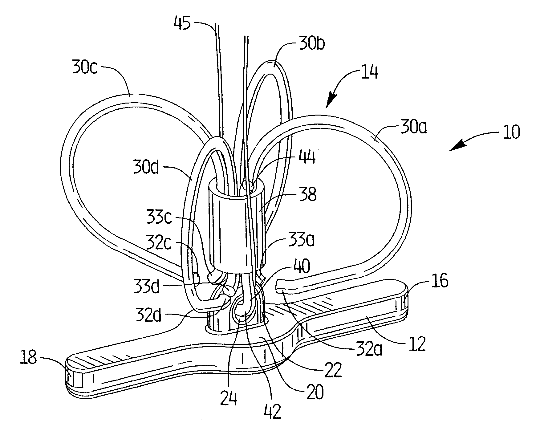
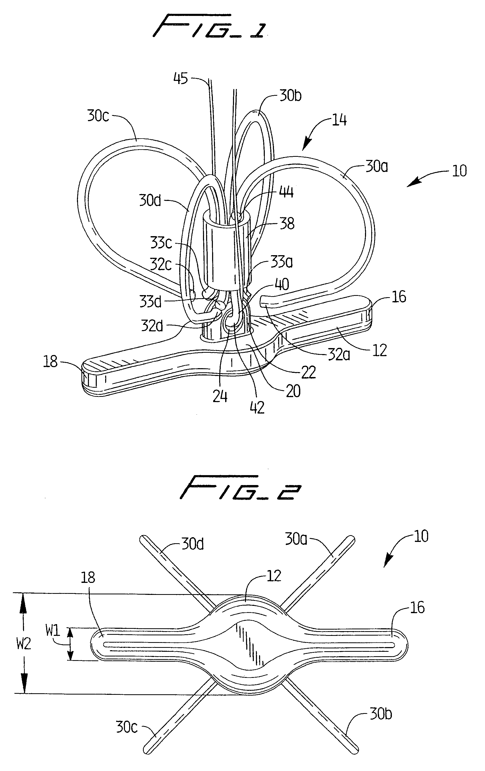
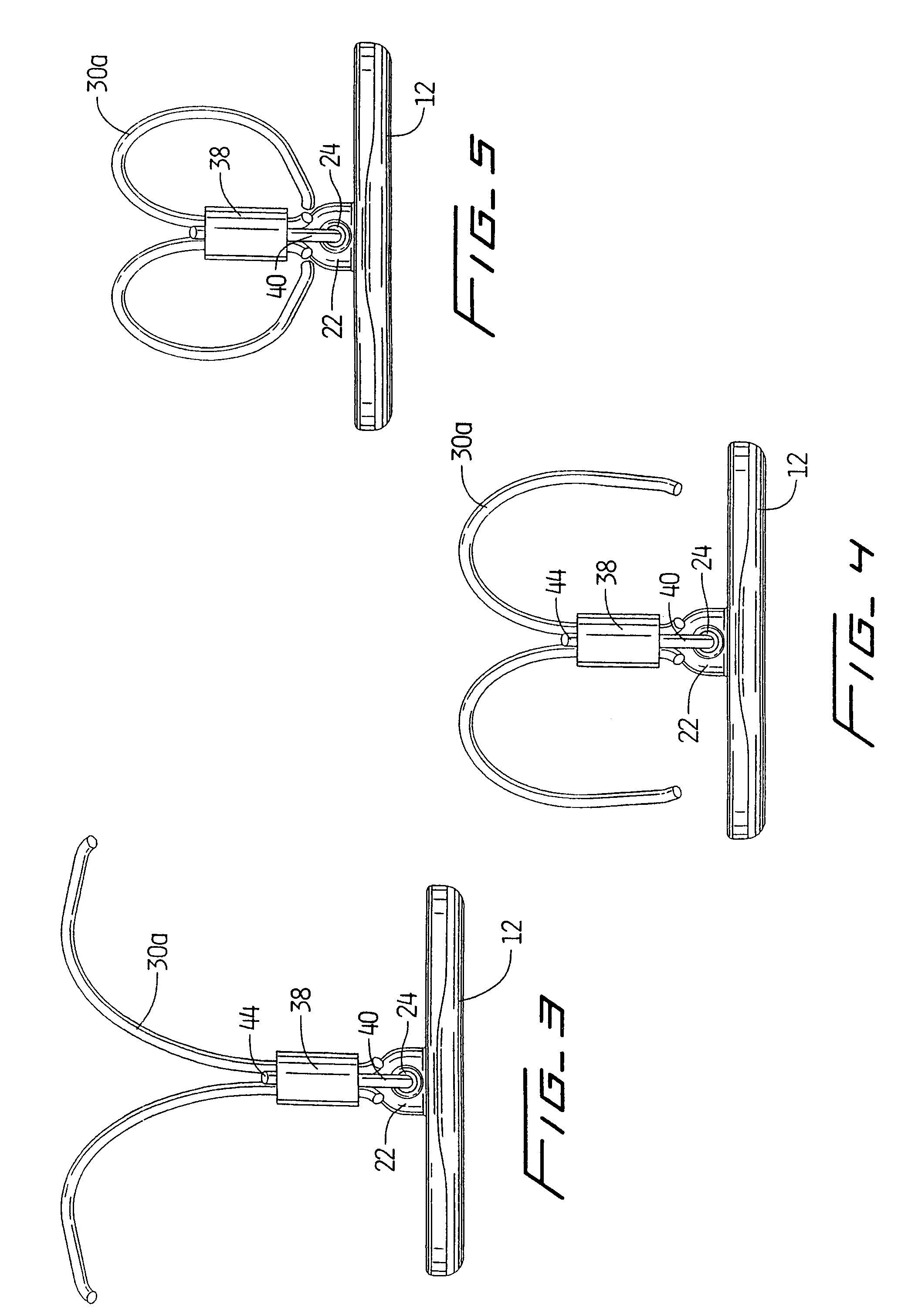
[0099]Referring now in detail to the drawings where like reference numerals identify similar or like components throughout the several views, FIG. 1 is a perspective view of first embodiment of the vascular hole (aperture) closure device of the present invention. The device is intended to close an aperture in the vessel wall, typically formed after removal of a catheter previously inserted through the vessel wall into the vessel lumen for performing angioplasty or other interventional procedures. The aperture extends through the patient's skin and underlying tissue, through the external wall of the vessel, through the wall of the vessel, and through the internal wall of the vessel to communicate with the internal lumen of the vessel. The closure devices of the present invention have a covering member or patch positioned within the vessel pressing against the internal wall of the vessel to block blood flow and a clip positioned external of the vessel wall to retain the covering membe...


PUM


Abstract

A device for closing an aperture in a vessel wall comprising an elongated member positionable inside the vessel against an internal opening of the aperture and having a dimension exceeding a dimension of the internal opening of the aperture to prevent egress of fluid through the aperture, and at least two curved legs positionable outside the vessel to help retain the elongated member in position.

Description

[0001]This application claims priority from provisional patent application Ser. No. 60 / 355,526, filed Feb. 6, 2002 and is a continuation-in-part of U.S. patent application Ser. No. 09 / 659,648, filed Sep. 12, 2000 now abandoned which claims priority from provisional patent application Ser. No. 60 / 153,736, filed Sep. 13, 1999.BACKGROUND[0002]1. Technical Field[0003]This application relates to a vascular device and more particularly to a device for closing openings in vessel walls.[0004]2. Background of Related Art[0005]During certain types of vascular surgery, catheters are inserted through an incision in the skin and underlying tissue to access the femoral artery in the patient's leg. The catheter is then inserted through the access opening made in the wall of the femoral artery and guided through the artery to the desired site to perform surgical procedures such as angioplasty or plaque removal. After the surgical procedure is completed and the catheter is removed from the patient, ...

Claims


Application Information

Patent Timeline
Patent Type & Authority Patents(United States)
IPC IPC(8): A61B17/08A61B17/00A61B17/064
CPCA61B17/0057A61B17/064A61B2017/00637A61B2017/00659A61B2017/00867
Inventor HINCHLIFFE, PETER W. J.MCGUCKIN, JR., JAMES F.BRIGANTI, RICHARD T.PETERS, WALTER H.
Owner REX MEDICAL LP
Learn More