Method and apparatus for acquiring digital microscope images

a digital microscope and image acquisition technology, applied in the field of digital microscope image acquisition, can solve the problems of time-consuming imaging process, difficult to diagnose the pathologist's image of the specimen, and difficulty in obtaining individual image tiles, so as to achieve the effect of reducing the processing time of image alignmen

Inactive Publication Date: 2008-06-17
SOURCE BIOSCI
View PDF13 Cites 22 Cited by
  • Summary
  • Abstract
  • Description
  • Claims
  • Application Information

AI Technical Summary

Benefits of technology

[0018]By acquiring field of view images whilst the microscope stage (and thus the specimen) is moving relative to the imaging system, in accordance with the present invention, it is possible to significantly reduce the imaging time for a complete specimen. The imaging system resource can therefore be used to image a greater number of specimens per day than would be imaged using conventional imaging techniques.

Problems solved by technology

Whilst such conventional methods of acquiring digital microscope images result in a high quality composite image of the specimen, the imaging process is time consuming, particularly if imaging of a complete specimen is performed using a medium or high power objective lens in the microscope.
Moreover, the time taken to acquire an individual image tile is subject to physical constraints.
This time delay in the image of the specimen becoming available for diagnosis by a pathologist is problematic.
In addition, the use of the valuable resource of the imaging system for such a lengthy period of time for only a single specimen may be difficult to justify.
However, this process represents a poor use of the pathologist's time.
In addition, if a remote pathologist is then consulted for a second opinion, the remote pathologist will not be able to view, at high magnification, areas of the specimen other than those selected by the local pathologist.
This compromises the ability of the remote pathologist to make an accurate diagnosis, since he or she is reliant on the local pathologist having selected all the important areas of the specimen.
Many consultant pathologists will be reluctant to give a diagnostic opinion based on the limited images selected by another pathologist, particularly if that pathologist is not know to him or her.
However, this solution has the disadvantage that there will be a further delay between the time the remote pathologist makes the selection and the time when high magnification images become available due to the time taken for the specimen area to be imaged.
However, as explained above, the process of acquiring adjacent high magnification images by stepwise movement of the stage is time consuming.

Method used


Image

  • Method and apparatus for acquiring digital microscope images
  • Method and apparatus for acquiring digital microscope images
  • Method and apparatus for acquiring digital microscope images

Examples


Embodiment Construction

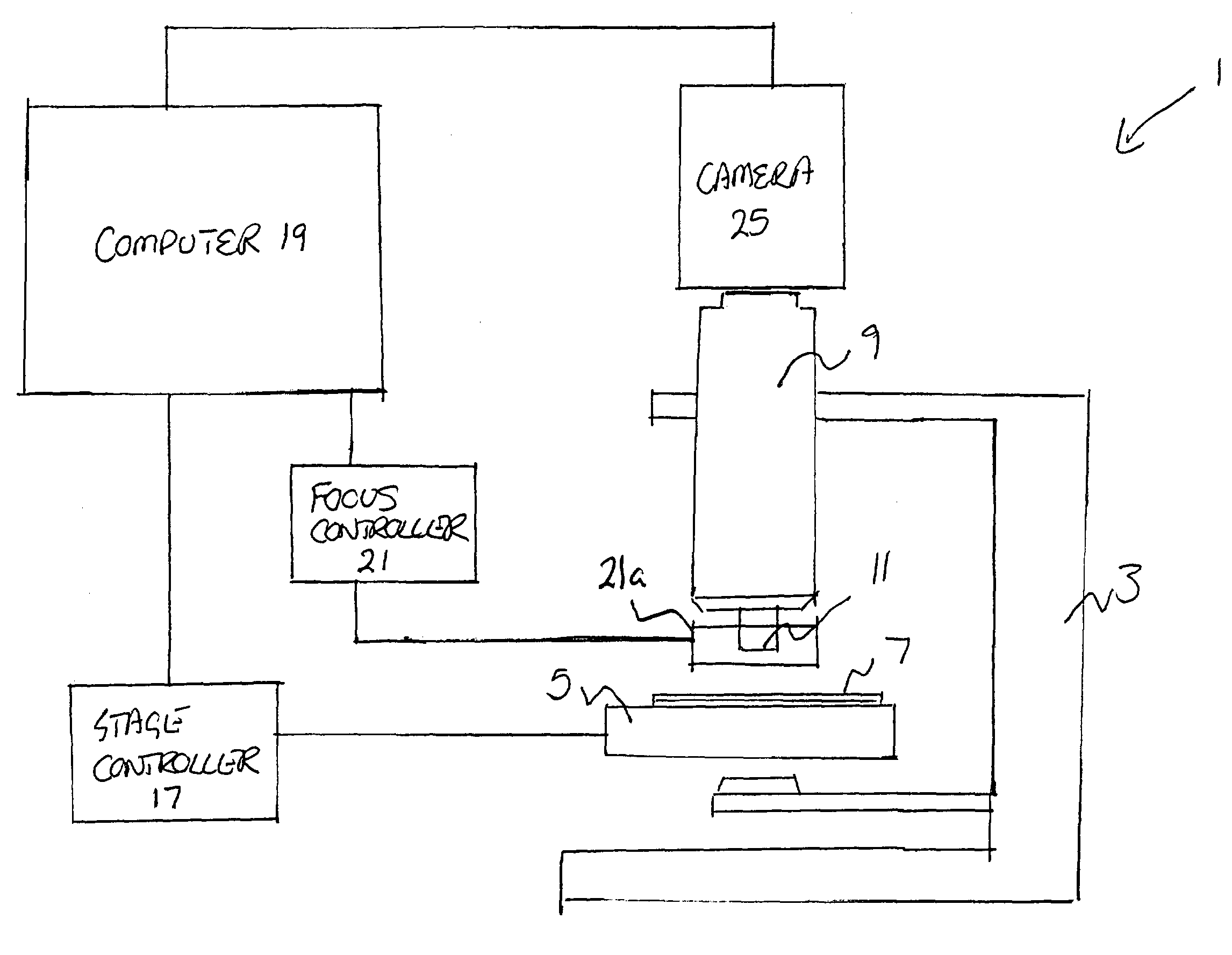
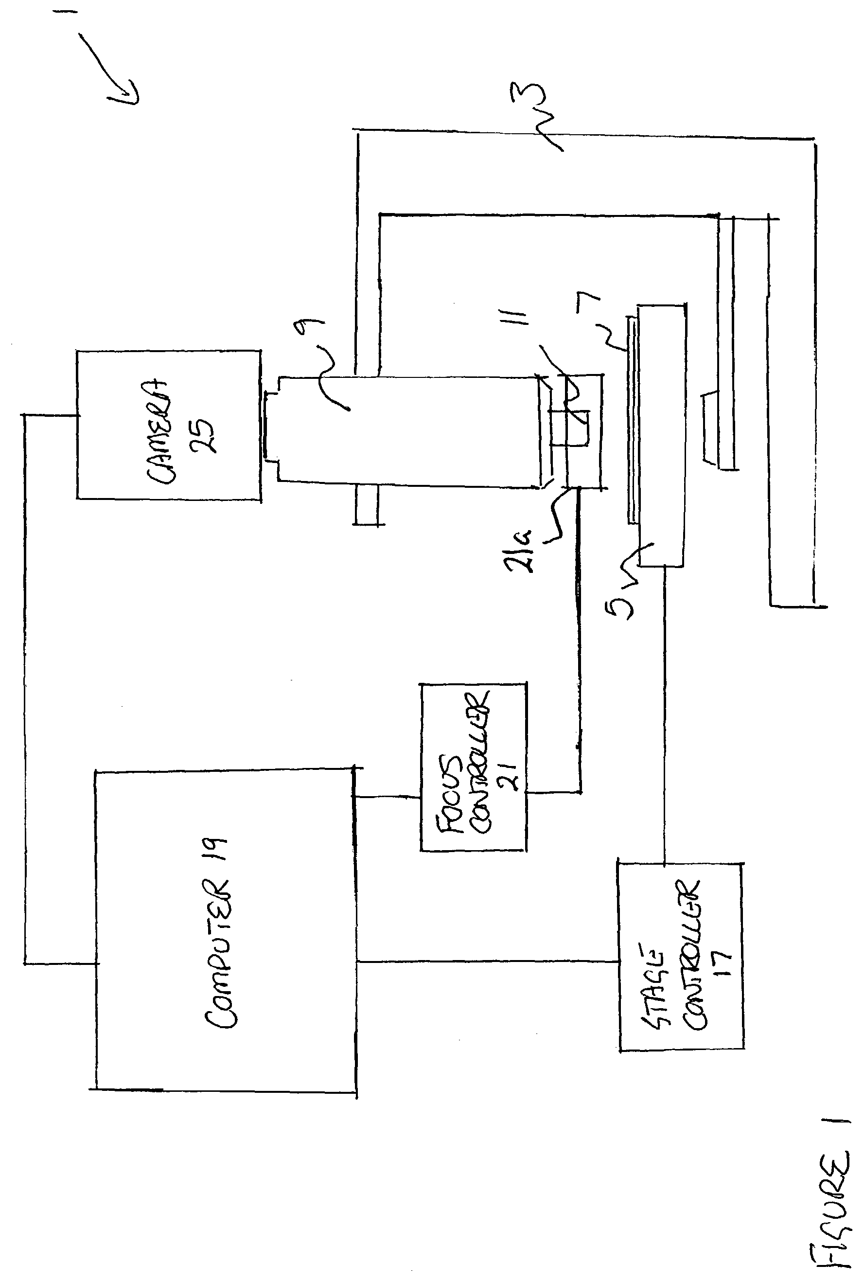
[0035]FIG. 1 illustrates the digital imaging apparatus suitable for use in accordance with a preferred embodiment of the present invention. The imaging apparatus 1 comprises a microscope 3 having a motorised microscope stage 5 for holding a microscope slide 7, and an optical subsystem 9 including an objective lens 11 and eyepiece 15. An example of a suitable microscope is the Axioplan 2 imaging microscope available from Carl Zeiss of Germany, using an Apochromat objective lens. The stage may be any suitable motorised stage having the necessary positioning accuracy, for example the Proscan stage available from Prior Scientific Instruments Limited of Cambridge, UK.

[0036]The motorised stage 5 is driven under the control of a stage controller 17, which controls the stage in response to instructions from computer 19. The motorised stage 5 is typically driven only in the x- and y- directions. In addition, focusing of the microscope is controlled by a piezo-electric controller 21 which con...


PUM


Abstract

A method and apparatus for acquiring digital microscope images is disclosed, in which a plurality of magnified images of a specimen are captured for tiling together to provide an overall composite image of the specimen. In accordance with the described method, the specimen is moved relative to an imaging system comprising a microscope and camera in a predetermined path whilst the plurality of magnified images are captured. In a preferred embodiment, the specimen, contained on a slide, is mounted on a movable microscope stage, and is moved beneath the microscope in the predetermined path. The velocity of the movement of the stage and the shutter speed of the camera is computer controlled to capture overlapping, clear images.

Description

PRIORITY CLAIM[0001]The present application claims priority to UK Patent Application No. 01 302 06.6, entitled “Method And Apparatus For Acquiring Digital Microscope Images”, which application was filed on Dec. 18, 2001.BACKGROUND OF THE INVENTION[0002]1. Field of the Invention[0003]The present invention relates to a method and apparatus for acquiring digital microscope images, and more particularly to a method and apparatus for acquiring a digital image of a specimen, or part thereof, on a microscope slide, which is larger than the field of view of the microscope.[0004]2. Description of the Related Art[0005]The use of digital images of microscope specimens, which are typically displayed on a display screen, is becoming increasingly important in the field of diagnostic pathology. For example, digital images of a microscope specimen may be captured using a microscopy imaging system and the acquired digital image data may be assembled together to form a composite image of the specimen...

Claims


Application Information

Patent Timeline
Patent Type & Authority Patents(United States)
IPC IPC(8): H04N7/18G02B21/36
CPCG02B21/367
Inventor MADDISON, JOHN RCLINCH, NOAH F
Owner SOURCE BIOSCI
Why Eureka
Try Eureka