
Fat/water separation and fat minimization magnetic resonance imaging systems and methods

a magnetic resonance imaging and fat/water separation technology, applied in the field of magnetic resonance imaging systems and methods, can solve problems such as errors in reconstructed images, foil quantitative measurements of small water signal features surrounded by fat-containing tissue, and object borders may also be corrupted by this artifa

Active Publication Date: 2006-08-29
GENERAL ELECTRIC CO
View PDF16 Cites 17 Cited by
  • Summary
  • Abstract
  • Description
  • Claims
  • Application Information

AI Technical Summary

Benefits of technology

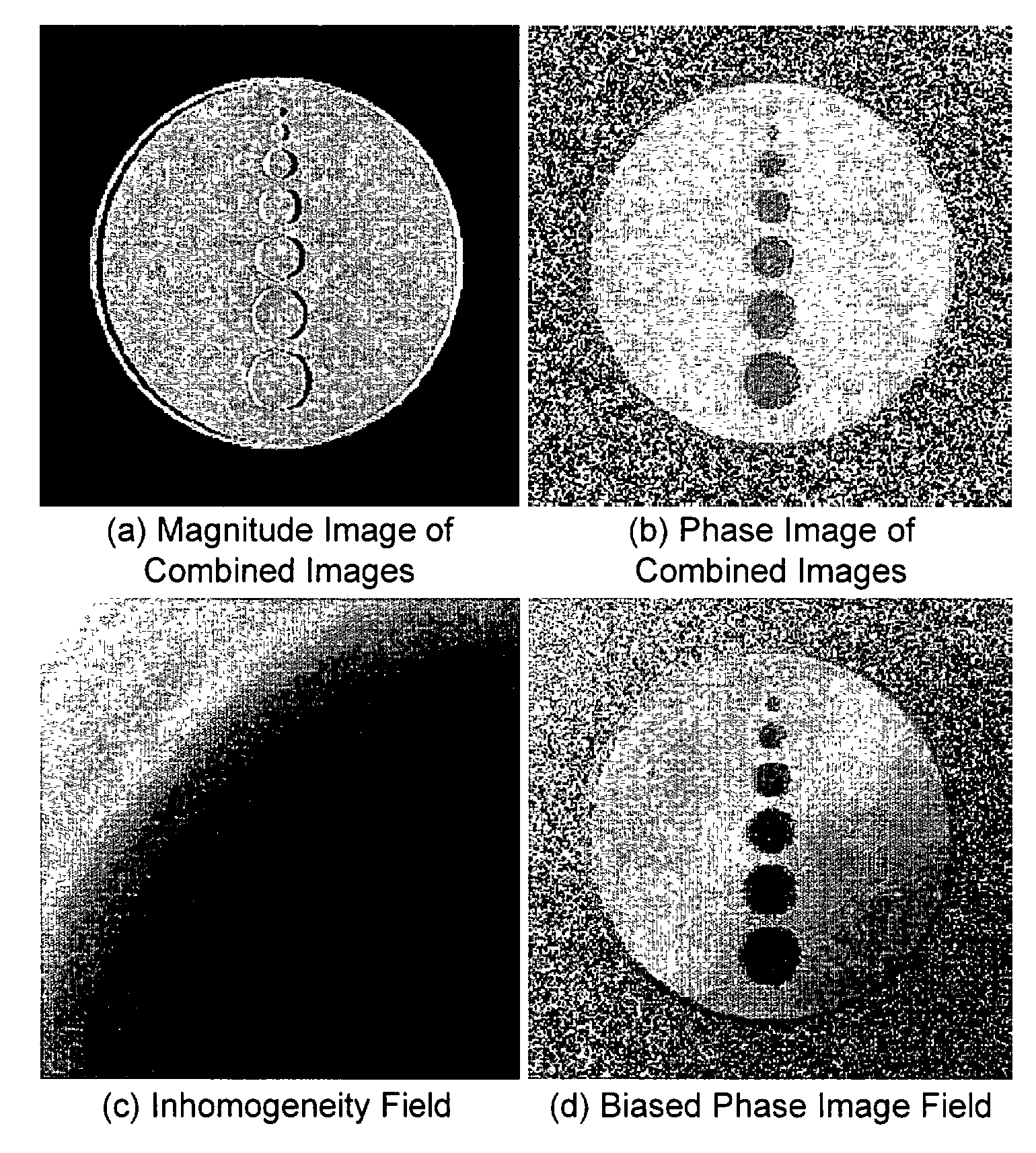
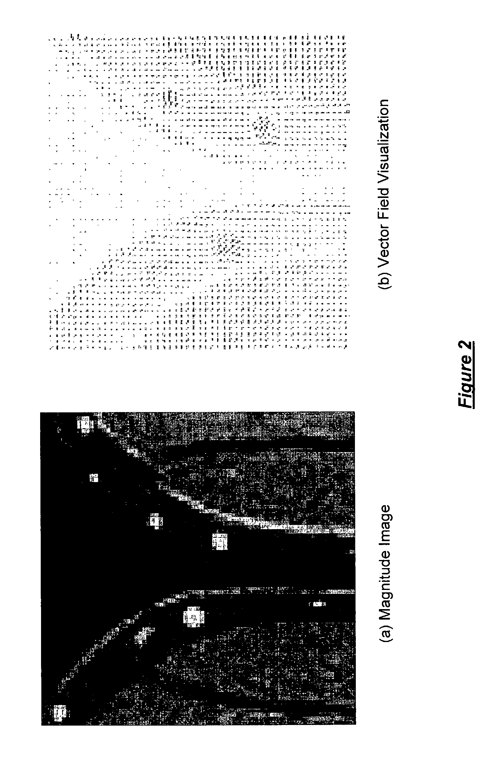
[0012]As described above, magnetic resonance imaging (“MRI”) fat-suppression techniques to overcome the chemical shift artifact (“CSA”) have been computationally approached primarily by variations of the Dixon method. Multi-point, and specifically, three-point, methods dominate the literature. These methods estimate the contribution of fat and water signals by solving a set of equations using three echoes acquired with the relative phase of fat and water at θε[−π, 0, π]. The systems and methods of the present invention, however, present a novel approach to fat suppression that requires only a single image acquisition. By choosing the relative phase to be approximately θ=π / 2 and employing an expectation maximization algorithm to classify the phase of the complex MR image, an estimate of the true phase of the image is obtained. From the phase estimate, fat and water images are obtained by projecting the complex vectors into fat and water components. The systems and methods of the present invention improve upon existing multi-point Dixon methods by reducing imaging time to a single image and eliminating the need for custom pulse sequence development. Mathematical and physical phantoms and clinically relevant results are reported.
[0017]In a further embodiment of the present invention, a method for minimizing a partial volume effect of fat and a chemical shift artifact associated with magnetic resonance (“MR”) imaging includes selecting a predetermined range for the phase inter-relationship between fat and water, selecting a phase inter-relationship value between fat and water within the predetermined range, and estimating a tissue parameter using the phase inter-relationship value between fat and water. Preferably, the predetermined range for the phase inter-relationship between fat and water includes values between about 60 degrees and about 120 degrees, and, more preferably, about π / 2. The tissue parameter estimated includes, for example, T1, T2, T2*, or diffusion.
[0018]In a still further embodiment of the present invention, a system operable for minimizing a partial volume effect of fat and a chemical shift artifact associated with magnetic resonance (“MR”) imaging includes an algorithm operable for selecting a predetermined range for the phase inter-relationship between fat and water, selecting a phase inter-relationship value between fat and water within the predetermined range, and estimating a tissue parameter using the phase inter-relationship value between fat and water. Preferably, the predetermined range for the phase inter-relationship between fat and water includes values between about 60 degrees and about 120 degrees, and, more preferably, about π / 2. The tissue parameter estimated includes, for example, T1, T2, T2*, or diffusion.

Problems solved by technology

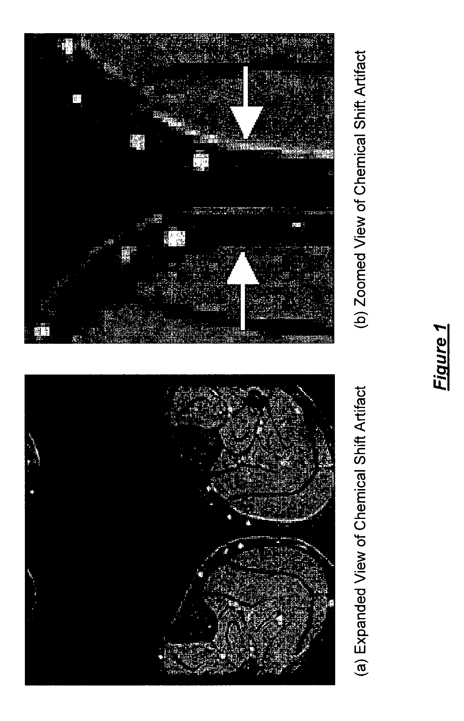
The chemical shift artifact (“CSA”) presents a significant barrier to the quantitative MR image analysis of small features, in which the amount of shift is comparable to the dimension of the object.
The borders of objects may also be corrupted by this artifact.
Typically, the resulting CSA corruption is “read through” by radiologists, however, by corrupting the borders between adjacent regions, it foils quantitative measurements of small water signal features surrounded by fat-containing tissue.
Intensity inhomogeneities, however, confound the two-point method, resulting in errors in the reconstructed images.
High bandwidth limits may alleviate this overlap, but increase noise.

Method used


Image

  • Fat/water separation and fat minimization magnetic resonance imaging systems and methods
  • Fat/water separation and fat minimization magnetic resonance imaging systems and methods
  • Fat/water separation and fat minimization magnetic resonance imaging systems and methods

Examples


working example

[0045]Images of two normal, healthy volunteers were acquired. One dataset was of the head and another was of the legs, as illustrated in FIG. 7. The head dataset was acquired using a GRE sequence on a 1.5 T GE Signa scanner with TE / TR of 6.1 msec / 700 msec and 42 degrees flip angle. Magnitude, real and imaginary images were saved by the scanner. The leg dataset was acquired on the same scanner with 9 msec / 117 msec TE / TR times and 20 degrees flip angle. The complex data was preprocessed using the phase unwrapping algorithm described above. The Guillemaud algorithm with φwater=φfat=0.2, Γwater=0, Γfat=mod2π(TE / 4.85 msec), and Γother=0.3. The results are illustrated in FIGS. 8 and 9.

[0046]The extended two-point Dixon method was implemented for comparison purposes, with the results illustrated in FIG. 10. No attempt was made to register the resulting images. Mis-registration artifacts may be seen in the “difference” images (FIGS. 10(c) and 10(f)). The single-point method provides qualita...


PUM


Abstract

Systems and methods for identifying the relative contribution of fat and water signals in a magnetic resonance (“MR”) image including an algorithm operable for selecting an image signal model, selecting a scan parameter, forming a bias field estimate, applying a bias correction to a phase image, estimating the signal fraction of fat and water at each of a plurality of voxels, and forming a fat-suppressed image, a water-suppressed image, or a combination of a fat-based image and a water-based image. The (“MRI”) fat suppression systems and methods requiring only a single image acquisition including an algorithm operable for selecting a relative phase of approximately θ=π / 2 or another suitable relative phase, employing an expectation maximization algorithm to classify the phase of the complex image, and projecting complex vectors into fat and water components to obtain fat and water images.

Description

FIELD OF THE INVENTION[0001]The present invention relates generally to magnetic resonance imaging (“MRI”) systems and methods. More specifically, the present invention relates to systems and methods for removing / reducing the chemical shift artifact associated with MRI using a single echo pulse sequence and for minimizing the partial volume effect of fat along an MRI slice select direction, as well as the chemical shift of fat along an MRI read direction.BACKGROUND OF THE INVENTION[0002]Magnetic resonance imaging (“MRI”) collects data in the Fourier domain, typically referred to as k-space, from the magnetic signals of protons precessing in a magnetic field. The spin frequency, also referred to as the resonance frequency, is a function of a material's gyromagnetic ratio and the strength of the magnetic field. In order to spatially position the collected signal, MRI systems and methods make an underlying assumption about this spin frequency: that the signal is composed primarily of pr...

Claims


Application Information

Patent Timeline
Patent Type & Authority Patents(United States)
IPC IPC(8): G06K9/00A61B5/055G01R33/483G01R33/54
CPCG01R33/4828G01R33/5607G01R33/56563
Inventor BLEZEK, DANIELDHAWALE, PARITOSHDIXON, WILLIAM
Owner GENERAL ELECTRIC CO
Learn More