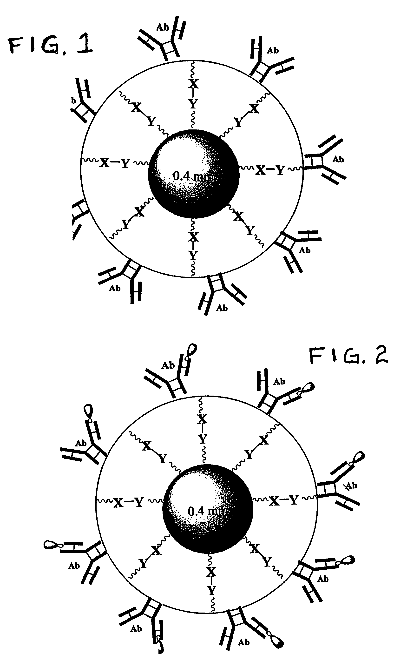
Beads for capturing target cells from bodily fluid
a technology of target cells and beads, which is applied in the field of beads for capturing target cells from bodily fluid, can solve the problems of greatly hindering the use of fetal cells from the circulation of mothers for diagnostic purposes, and the fact that fetal cells are present in maternal blood in only very limited numbers, and achieves excellent sequestering agents, high porousness, and efficient separation or isolation
- Summary
- Abstract
- Description
- Claims
- Application Information
AI Technical Summary
Benefits of technology
Problems solved by technology
Method used
Image
Examples
example 1
[0059]Polystyrene beads were obtained from Duke Scientific Corporation which are spheroids that have diameters of about 200 microns. The beads are not otherwise surface-treated by the manufacturer and, as a result, are hydrophobic. The beads (0.5 g) were first subjected to plasma activation and then aminated with 50% triethoxysilylpropaneamine in 5 ml of dry CH2Cl2. The beads were then washed with ethanol (3×2 ml) and water (3×2 ml).
[0060]The aminated beads were mixed with a solution containing 3 weight % of a hydrogel prepolymer plus a selective antibody of choice. For example, an initial solution is made containing about 12.5 milligrams of a prepolymer; the prepolymer is made by reacting PEG having a MW of about 6000 daltons, TMP and IPDI (about 0.15 mole TMP:about 3.1 moles IPDI; about 1 mole PEG) at 120° C. under dry nitrogen in the absence of a catalyst. The resultant urethane prepolymer is isocyanate-terminated and has a reactive isocyanate content of about 0.4 meq. per gram. ...
example 2
[0061]In order to produce satisfactory beads made entirely of hydrogel, a large crystalizing dish is filled with mineral oil, and a stir bar is set for 200 rpm. About 100 μl of a solution similar to that described in Example 1, containing about 3% of the prepolymer but instead containing 4 μl (1 μg) of a CD71 antibody at a concentration of about 0.25 mg / ml, is fed through a syringe pump for discharge into the oil bath through a needle having a opening of about 100 microns ID. The pump is operated to create a steady rate of flow of the aqueous solution exiting the orifice, e.g. 1 milliliter per minute (1 ml / min).
[0062]The velocity of the oil that is caused to move past the needle (by the action of the stir bar as described above) and the ID of the injection needle determine the size of the hydrogel droplets that break off at the tip of the needle. As each droplet is broken off, it moves away from the needle, and by using a sufficiently large crystalizing dish, the beads that are form...
example 3
Use of the Coated Beads of Example 1 to Isolate Specific Cells
[0063]A cell suspension of cells from cervical mucous substantially the same as that prepared in Comparative Example 5 was prepared. About 5 mg of B1 beads with the Trop 2 Abs bonded thereto were incubated with the cell suspension in a total volume of about 250 μl for about 1 hour at 4-8° C. with gentle mixing. The beads were washed with PBS / BSA (3×200 μl) and analyzed under microscope. About 95% of the trophoblasts were captured on Trop 2-coated beads showing a very substantial improvement upon the performance of the CB#3C beads which carried the same Abs.
PUM
| Property | Measurement | Unit |
|---|---|---|
| size | aaaaa | aaaaa |
| size | aaaaa | aaaaa |
| molecular weight | aaaaa | aaaaa |
Abstract
Description
Claims
Application Information
 - R&D
- Intellectual Property
- Life Sciences
- Materials
- Tech Scout
- Unparalleled Data Quality
- Higher Quality Content
- 60% Fewer Hallucinations
Browse by: Latest US Patents, China's latest patents, Technical Efficacy Thesaurus, Application Domain, Technology Topic, Popular Technical Reports.
© 2025 PatSnap. All rights reserved.Legal|Privacy policy|Modern Slavery Act Transparency Statement|Sitemap|About US| Contact US: help@patsnap.com

