Methods and systems for measuring the size and volume of features on live tissues

a technology of live tissues and features, applied in the field of size and volume measurement, can solve the problems of major source of inaccuracy or variability, obvious pitfalls of the instrument and methodology used to calculate volume as well as operator-related errors, and approach is fairly time-consuming

Active Publication Date: 2008-11-11
BIOPTICON CORP
View PDF13 Cites 30 Cited by
  • Summary
  • Abstract
  • Description
  • Claims
  • Application Information

AI Technical Summary

Benefits of technology

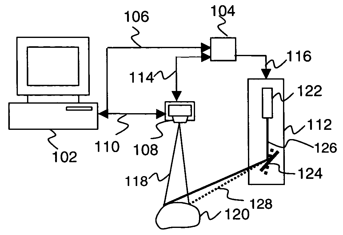
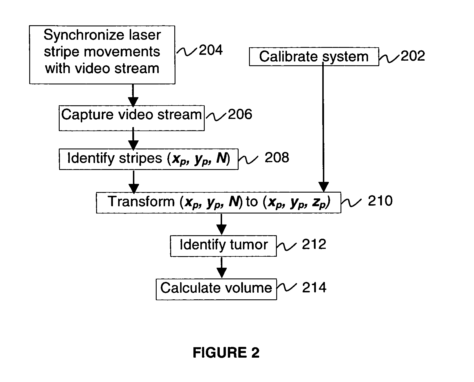
[0010]The methods and systems of the present invention utilize a 3D structured light scanner and geometry based feature recognition logic to obtain shape, size and / or volume of the animal surface features observed in clinical studies. Such features may include tumors, lesions, sores, and wounds and any other suitable feature on the surface of animals, plants and humans, and any other suitable surface. The invention may obtain a number of 2D images of the surface feature, such as a tumor, with patterns of structured light projected on the feature. The parallax between a camera and a light source is used to compute a 3D point cloud of the feature's surface. The point cloud is a collection of (x, y, z) coordinates of the surface feature points. Recognition algorithm separates the feature points from the healthy points by minimizing the standard deviation of the point cloud points from the suggested shape. After the separation is achieved, the feature size and volume may be calculated.
[0012]In this way, the invention provides for fast, non-contact, automated measurements of external features such as tumors, lesions, sores, wounds shape, size and volume on the surface of animals, plants and humans.
[0013]The invention also provides time savings, which are extremely important when measuring, for example, tumor progress using large animal sets. For example, the 3D scanner of the present invention is many times faster than CAT methods. Compared to caliper method, the 3D scanner measurements are also faster. In addition, the 3D scanner method may be completely automated (i.e., measurement data may be electronically transferred to a computer from a measuring device, eliminating extra time needed to record and transfer paper recorded data to computers).
[0014]The invention may further be totally non-invasive (i.e., no part of the device touches the animal). This leads to three important consequences, for example, for making tumor volume measurements: i) animals are not stressed as much, which less affects the tumor growth; ii) there is no need to sterilize the device; and iii) tumors are not deformed by the measuring equipment. Only the 3D scanner light touches the animals. The 3D scanner method does not involve injecting contrast agents or radioactive substances into animals. Since it takes a very short time, around a few seconds, to complete the measurement, the 3D scanner method is less stressful to animals and preferably does not require anesthetizing them.
[0016]Further in accordance with the invention, certain embodiments also include: basing the separation and calculation upon the feature portion of the surface having a predetermined feature shape and the non-feature portion of the surface having predetermined non-feature shape; the: predetermined feature shape and the predetermined non-feature shape being described by a finite set of parameters; performing successive approximations to find the set of parameters; selecting an arbitrary initial choice of the set of parameters for the successive approximations; calculating distances between coordinates in the set and each of the predetermined feature shape and the predetermined non-feature shape; assigning the coordinates in the set to the feature portion of the set when the coordinates are closer to the predetermined feature shape than to the predetermined non-feature shape, and to the non-feature portion of the set when the coordinates are closer to the predetermined non-feature shape than to the predetermined feature shape; finding the set of parameters by minimizing the standard deviation of the feature portion of the set from the predetermined feature shape and by minimizing the standard deviation of the non-feature portion of the set from the predetermined non-feature shape; and calculating the size of the predetermined feature shape based on the set of parameters and estimating the size of the feature portion of the surface to be the size of the predetermined feature shape.

Problems solved by technology

Unfortunately, this approach has obvious pitfalls related to the instrument and methodology used to calculate volume as well as operator-related errors.
First, a major source of inaccuracy or variability is caused by the operator's judgment on the choice of the ellipsoid axes.
Second, this approach is fairly time consuming.
In the case of cancer studies, for example, a single study may involve hundreds of animals, and because the tumor volume measurements have to be done at least twice a week and take a given amount of investigator time per measurement, there is a limit in the number of studies that a single investigator can handle at once.
Such devices require direct contact to features such as tumors, however.
The use of such methods is not feasible in many cases, however, due to long and costly preparation times, the unwanted effects of the contrast agents and radioactive compounds and the necessity to anesthetize animals during measurement.
Notwithstanding the availability of the foregoing techniques, there is no method or device for non-contact and fast measurement of the volume of surface features of small animals that can be used in, for example, pre-clinical cancer and inflammation studies.
For example, manual caliper measurements are prone to significant subjective operator errors; CAT, PET, MRI and SPECT methods are unacceptably time consuming and expensive; and caliper and cylindrical chamber methods require direct contact with the object under measurement, which is a potential source of feature deformation and contamination that may negatively effect the measurements being made.

Method used


Image

  • Methods and systems for measuring the size and volume of features on live tissues
  • Methods and systems for measuring the size and volume of features on live tissues
  • Methods and systems for measuring the size and volume of features on live tissues

Examples


Embodiment Construction

[0023]In the following detailed description, numerous specific details are set forth regarding the methods and systems of the present invention and the environments in which the methods and systems may operate in order to provide a thorough understanding of the present invention. It will be apparent to one skilled in the art, however, that the present invention may be practiced without such specific details. In other instances, well-known components, structures and techniques have not been shown in detail to avoid unnecessarily obscuring the subject matter of the present invention. Moreover, various examples are provided to explain the operation of the present invention. It should be understood that these examples are merely illustrative and various modifications may be made in accordance with the invention. It is contemplated that there are other methods and systems that are within the scope of the present invention.

[0024]Many designs of the 3D structured light scanners are known, ...


PUM


Abstract

Methods and systems for measuring volume and size of features such as lesions, tumors, sores, and wounds external to animal, human and plant skin, using 3D structured light scanners are provided. These methods and systems obtain a point cloud of the scanned feature, and employ algorithms to adjust the suggested feature geometry parameters to minimize deviations of the point cloud from suggested feature geometry. Obtained geometry parameters permit the calculation of the features' sizes and volumes.

Description

CROSS REFERENCE TO RELATED APPLICATION[0001]This application claims the benefit under 35 U.S.C. § 119(e) of U.S. Provisional Patent Application No. 60 / 502,364, filed Sep. 12, 2003, which is hereby incorporated by reference herein in its entirety.FIELD OF THE INVENTION[0002]This invention relates generally to the field of measurement of size and volume, and, in particular, to the measurement of size and volume of features such as tumors, lesions, sores, wounds on the surface of animals, plants and humans using 3D image construction from 2D images of the object and patterned light superimposed over the object.BACKGROUND[0003]The identification and selection of potentially active drugs for use in treatment of diseases, (for example, cancer) has largely been based on the screening of large numbers of compounds and rational drug design (when the molecular structure of the target such as tumor is known). Once compounds have been chosen based on selectivity to the target and desired functi...

Claims


Application Information

Patent Timeline
Patent Type & Authority Patents(United States)
IPC IPC(8): G06K9/36G06K9/00A61B5/103A61B5/107G01BG01B11/25
CPCA61B5/0062A61B5/0064A61B5/107A61B5/444A61B5/445G01B11/2518G06T7/0057Y10S128/916Y10S128/923G06T7/521
Inventor TALAPOV, ANDREI L.GIRIT, IBRAHIM CEM
Owner BIOPTICON CORP
Why Eureka
Try Eureka