Breast compression for digital mammography, tomosynthesis and other modalities

a technology of digital mammography and compression breast, applied in the field of breast compression, can solve the problems of not being transparent to visible light, the compression force is not uniformly distributed throughout the breast, and the need no longer compelling, so as to improve patient comfort in breast compression, and compress the thicker part of the breast mor

Active Publication Date: 2009-02-10
HOLOGIC INC
View PDF27 Cites 35 Cited by
  • Summary
  • Abstract
  • Description
  • Claims
  • Application Information

AI Technical Summary

Benefits of technology

[0003]A significant patient concern in mammography is the discomfort the patient feels when the breast is compressed, typically between two rigid plastic surfaces, with sufficient force to spread out the breast tissues. The reasons for using such high compression include: (1) to make the breast thinner in the direction of x-ray flux and thereby reduce patient radiation exposure from the level required to image the thicker parts of an breast that is not compressed; (2) to make the breast more uniform in thickness in the direction of x-ray flux and thereby facilitate more uniform exposure at the image plane over the entire breast image; (3) to immobilize the breast during the x-ray exposure and thereby reduce image blurring; and (4) to bring breast tissues out from the chest wall into the exposure area and thus image more tissue.
[0006]Some compression paddles tilt, such as the paddle available in various sizes from Lorad of Danbury, Conn., a division of Hologic, Inc. of Bedford, Mass., under the trade name F.A.S.T. This tilting paddle provides more uniform compression across the breast, and more comfortable breast examinations. An example of such a paddle is illustrated in FIG. 11, as mounted in a mammography system commercially available from Lorad. It is illustrated in FIG. 12 as used to compress a patient's breast in a similar mammography system.
[0012]Another device that can be used to improve patient comfort in breast compression is a similar pillow or bag but filled at least partly with a liquid such as water rather that filled only with a gas such as air. In addition to the benefits of a gas-filled device discussed above, the device that is filled at least partly with a liquid such as water would provide some equalization of the x-ray path length that forms the breast image.
[0013]Another device for improving patient comfort in breast compression is a sheet of transparent and sufficiently strong material such as Mylar to take over some or all of the functions of a compression paddle. The material may be supported by rods or other holders that flank the breast and are supported and are movable in a manner similar to that of a compression paddle. Alternatively, the material may be supported between rods or other holders that are secured to the breast platform or another part of the mammography system, in which case the need for a compression paddle may be eliminated altogether. The material may be stretchable or otherwise deformable in ways that are not uniform across the portion thereof used to compress the breast. For example, one or more portions, or all the material, closer to the chest wall may stretch or deform less that portions or all the material further from the chest wall of the patient, to thereby compress the thicker part of the breast more. In addition, at least some portions of the material facing the breast may be made sticky, for example by making the material itself sticky or by coating it with a sticky substance, in order to eliminate or reduce slippage between the breast and the material.
[0014]Yet another device that improves comfort in breast compression is made wholly or in part of a compressible foam or similar material. The device may have substantially constant thickness in the direction of x-ray flux, or it may be shaped to provide a shallow depression that cradles the breast.

Problems solved by technology

With the advent of digital mammography and its wide dynamic range, this requirement is no longer as compelling, because image processing can flatten the image post-exposure breast thickness.
One reason for the discomfort felt using this method is that the compression force is non-uniformly distributed throughout the breast.
Another pad system is proposed in U.S. Pat. Nos. 6,850,590 and 6,975,701 and published U.S. patent applications US 2006 / 0050844 A1, US 2004.0156472 A1 and US 2003 / 0099325 A1, all naming as the inventor Benjamin M. Galkin. One disadvantage of pad systems of this type is that the pads are not transparent to visible light.
This means that there is impaired visibility of the breast, if the pad is used above the breast.
Another disadvantage of this solution is that the pad needs to be made of fairly dense thin form, so as to provide at least meaningful deformability under the high pressures.
It is believed that this foam can creates minor artifacts in the image, especially if the pad slips during positioning and does not cover the entire breast.
Another limitation is that this pad is relatively expensive to manufacture, and this makes it more expensive to use, and that it may require excessive time to secure to and then remove from the breast platform and / or compression paddle.
However, newer modalities such as digital mammography and tomosynthesis have different needs and constraints, offer new challenges and new opportunities, and have allowed for reconsideration the old approaches.
In addition, no indication has been found in those proposals to make the pads transparent to visible light and thereby facilitate breast positioning in the compression process.
The material may be stretchable or otherwise deformable in ways that are not uniform across the portion thereof used to compress the breast.

Method used


Image

  • Breast compression for digital mammography, tomosynthesis and other modalities
  • Breast compression for digital mammography, tomosynthesis and other modalities
  • Breast compression for digital mammography, tomosynthesis and other modalities

Examples


Embodiment Construction

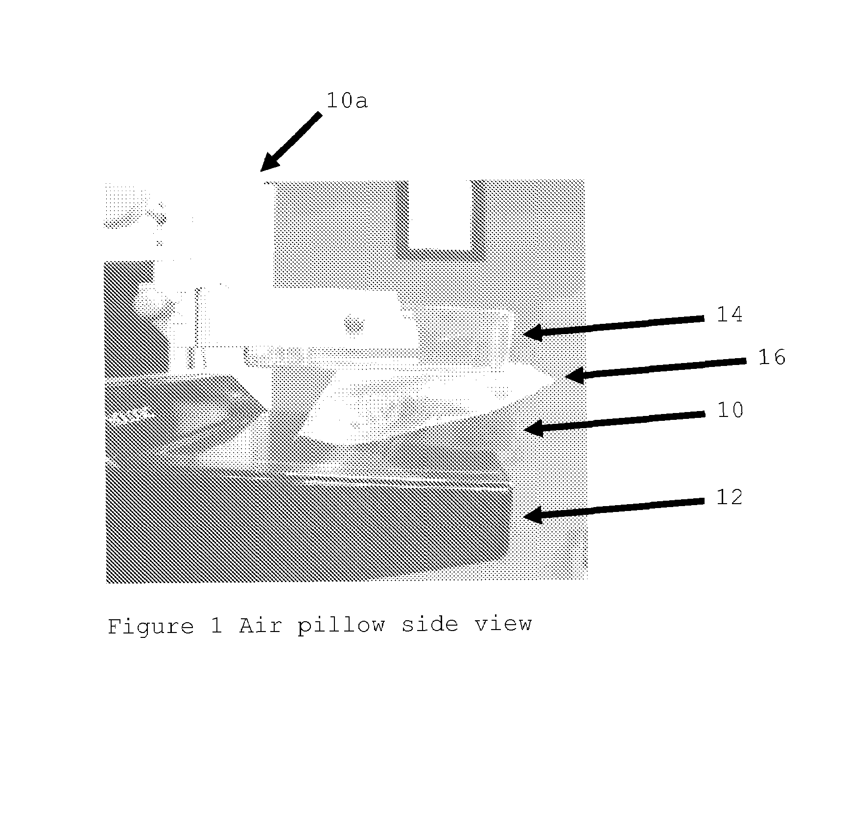
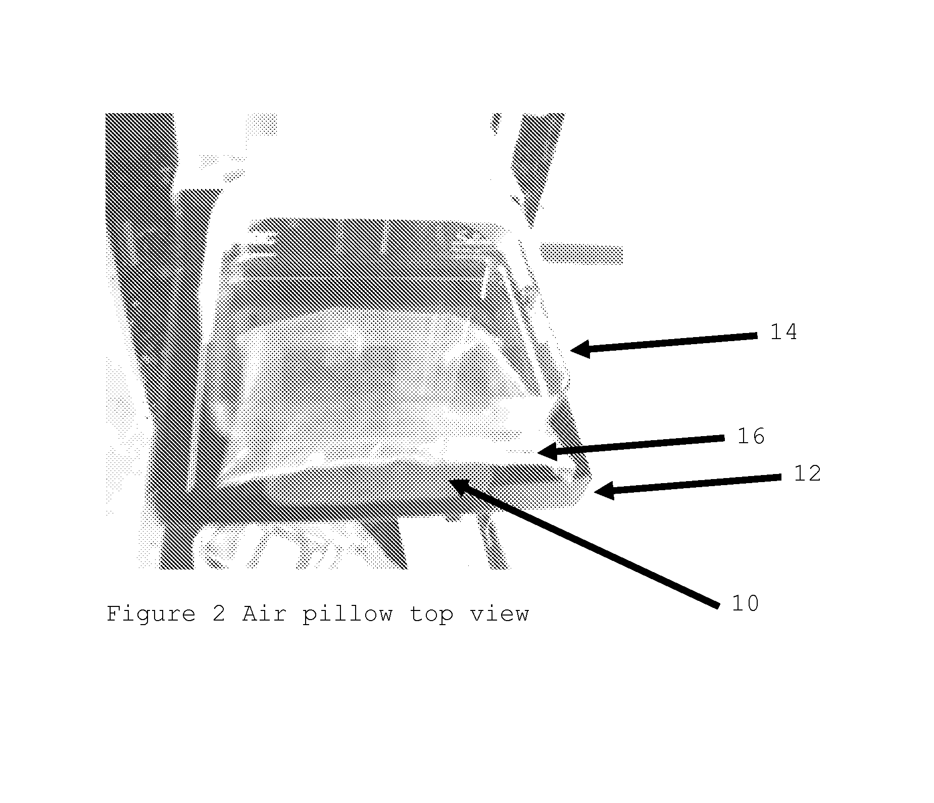
[0029]FIG. 1 illustrates a breast phantom 10 positioned for x-ray examination in a mammography or tomosyntesis system generally indicated at 10a. One example of a system 10a is the digital mammography system offered for sale currently by the Lorad division of Hologic, Inc. under the designation Selenia. Phantom 10 is between a breast platform 12 and a compression paddle 14. A device 16, in the form of a gas-filled bag or pillow, is placed between the breast and the compression paddle. In actual use, the patient would be sitting or standing to the right of breast platform 12 for an x-ray image taken at the CC orientation, with a breast 10 in place of phantom 10. The patient's chest wall would be generally along the vertical. FIG. 2 illustrates in perspective view the same arrangement, and FIG. 3 illustrates the device as it is before being compressed between the breast and the compression paddle. A similar device can be compressed between breast 10 and breast platform 12 in addition ...


PUM


Abstract

A breast x-ray imaging method and system that is particularly suited for tomosynthesis imaging but also is useful for conventional mammography. A fluid containing pillow or bag is placed between the breast and a paddle that compresses the breast against a breast platform covering an imaging device, to enhance patient comfort and provide other benefits. Alternatives include a flexible sheet compressing the breast, and a compressible foam, preferably contoured to accommodate a patient's breast.

Description

CROSS-REFERENCE TO RELATED APPLICATION[0001]This disclosure claims the benefit of U.S. Provisional Application Ser. No. 60 / 786,529, filed Mar. 27, 2006, the entire contents of which are incorporated by reference herein.FIELD[0002]This patent specification pertains mainly to mammography and finds particular application in digital mammography and tomosynthesis.BACKGROUND AND SUMMARY OF THE DISCLOSURE[0003]A significant patient concern in mammography is the discomfort the patient feels when the breast is compressed, typically between two rigid plastic surfaces, with sufficient force to spread out the breast tissues. The reasons for using such high compression include: (1) to make the breast thinner in the direction of x-ray flux and thereby reduce patient radiation exposure from the level required to image the thicker parts of an breast that is not compressed; (2) to make the breast more uniform in thickness in the direction of x-ray flux and thereby facilitate more uniform exposure at...

Claims


Application Information

Patent Timeline
Patent Type & Authority Patents(United States)
IPC IPC(8): A61B6/04H05G1/00
CPCA61B6/0414A61B6/502A61B6/025
Inventor DEFREITAS, KENNETHHOPPONEN, LINDASHAW, IANNIKLASON, LORENSTEIN, JAYSMITH, ANDREW P.
Owner HOLOGIC INC
Why Eureka
Try Eureka