Method and device for producing high-quality fundus images

a high-quality, fundus technology, applied in the field of high-quality fundus images, can solve the problems of inhomogeneity in the sensitivity of pixels, decrease in the resolution of color sensors, and complicating the creation of high-resolution color images, so as to improve image sharpness, reduce noise, and improve resolution

Inactive Publication Date: 2012-07-05
CARL ZEISS MEDITEC AG
View PDF6 Cites 32 Cited by
  • Summary
  • Abstract
  • Description
  • Claims
  • Application Information

AI Technical Summary

Benefits of technology

[0017]The present invention addresses the problem of providing a solution for producing high-quality fundus images which eliminates the disadvantages of the solutions of prior art and with which fundus images with higher resolution and dynamic as well as improved image sharpness and decreased noise can be realized at a lower radiation exposure of the patient's eye.

Problems solved by technology

CMOS sensors are most notably less expensive but not as light-sensitive as CCD sensors and frequently exhibit distinct inhomogeneities in the sensitivity of the pixels.
This decrease in resolution of the color sensors additionally complicates the creation of high-resolution color images of the retina and therefore also an exact diagnosis.
However, experience has shown that an increase of the number of color pixels through combining 4 pixels of the Bayer pattern leads only theoretically to a higher resolution because in the field, no significant increase of the resolution was detected.
A significant disadvantage of such sequential methods is the merging and / or superimposing of the monochromatic images to a color image through an algorithm.
This requires a calculation-intensive image postprocessing which is error-prone to some extent because the features are frequently not present in an exactly reproducible fashion.
This negatively affects the resolution as well as the image quality.
A further disadvantage of such a method is the relatively long exposure time of such a sequence of three monochromatic images.
This poses a significant disadvantage.
However, disadvantageously, disruptive influences of the measuring arrangement, e.g., the noise of the image acquisition sensor, still negatively affect the measurement results.

Method used


Image

  • Method and device for producing high-quality fundus images
  • Method and device for producing high-quality fundus images
  • Method and device for producing high-quality fundus images

Examples


Embodiment Construction

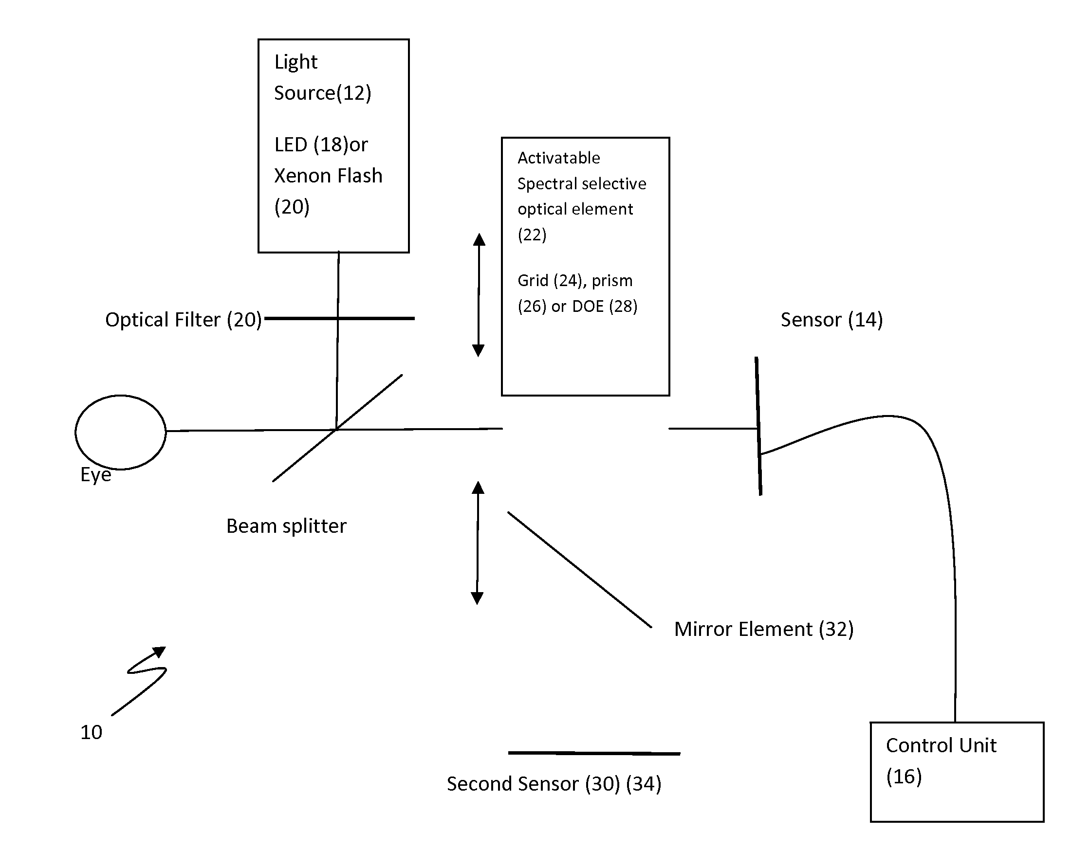
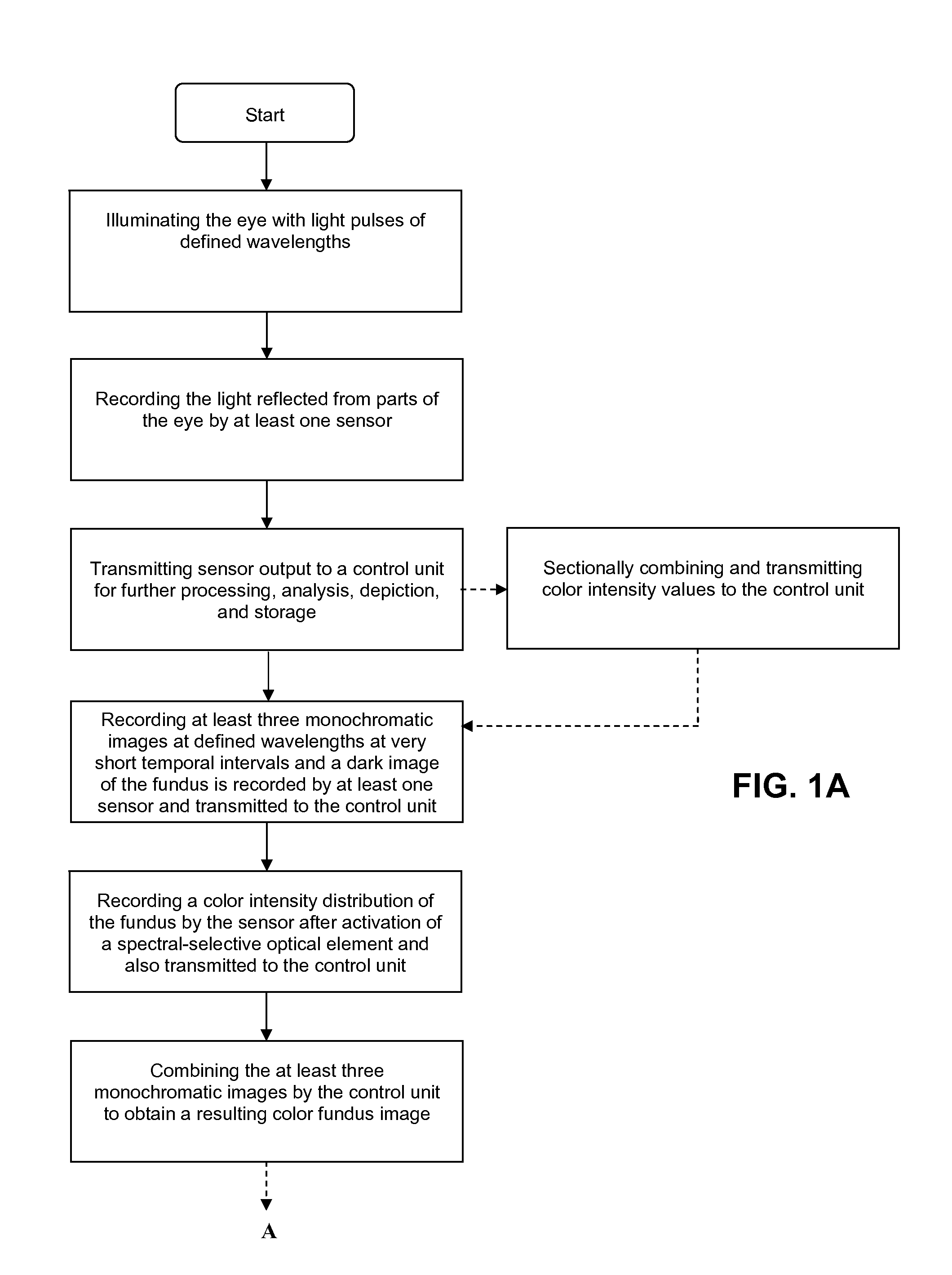
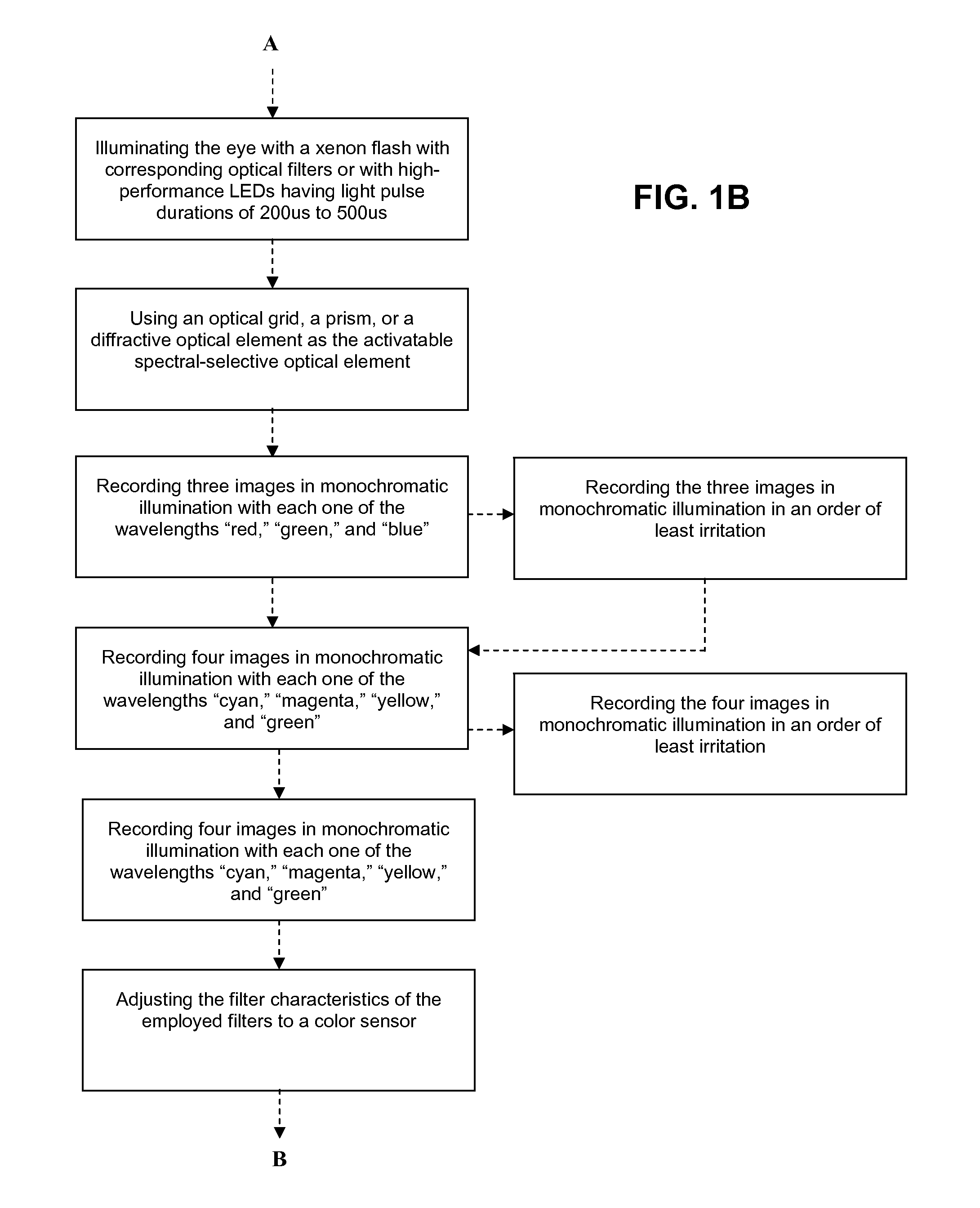
[0054]With the method for producing high-quality fundus images, according to the invention, the eye is illuminated with light pulses of defined wavelengths, the light reflected from parts of the eye is recorded by at least one sensor and transmitted to a control unit for

further processing, analysis, depiction, and storage. Thereby, at least three monochromatic images at defined wavelengths are recorded in very short temporal intervals, and subsequently a dark image of the fundus is recorded by at least one sensor and transmitted to the control unit. After activation of a spectral-selective optical element, the sensor records a color intensity distribution of the fundus at white illumination, which is also transmitted to the control unit. The at least three monochromatic images are combined by the control unit to obtain a resulting color fundus image, wherein the color intensity distribution is used for the correction of the color composition and the dark image is used for taking int...


PUM


Abstract

To produce a color fundus image, the eye is illuminated with light pulses of defined wavelengths. Light reflected is recorded by a sensor and transmitted to a control unit. At least three monochromatic images at very short temporal intervals and a dark image of the fundus are recorded. After activation of a spectral-selective optical element, a color intensity distribution of the fundus is recorded by the sensor at white illumination. The monochromatic images are combined by the control unit to obtain a resulting color fundus image, wherein the color intensity distribution is used for the correction of color composition and the dark image is used for taking into account the noise of the sensor. The solution permits monitoring, documenting and/or diagnosing of the fundus and can also be executed with ophthalmological systems based on the principle of optical coherence and/or confocal imaging.

Description

FIELD OF THE INVENTION[0001]The present invention relates to a method and a device for producing high-quality images of the fundus of an eye for later diagnosis through analysis of chromatic high-resolution images of the fundus.BACKGROUND[0002]While, according to prior art, slit lamps or laser scanning ophthalmoscopes are, e.g., used for images of the entire eye structures, fundus cameras have become generally accepted for images of the fundus.[0003]A fundus camera can map a precise image of the eyeground and thereby particularly depict the retina, the blood vessels, the optic nerve, and the choroid. It is used for diagnosis and for tracking the progress of diseases. Generally, the pupil of the patient must be dilated with medication when a fundus camera is used. It is known from prior art that no pupil reaction of the patient occurs in case of illumination of the eyeground by means of infrared light (invisible), therefore, a dilation of the pupil in a dark room occurs without the u...

Claims


Application Information

Patent Timeline
Patent Type & Authority Applications(United States)
IPC IPC(8): A61B3/14
CPCA61B3/14A61B3/12
Inventor MOHR, THOMASRO BERGER, BJORN
Owner CARL ZEISS MEDITEC AG
Why Eureka
Try Eureka