Non-invasive device for synchronizing chest compression and ventilation parameters to residual myocardial activity during cardiopulmonary resuscitation

a non-invasive, cardiac resuscitation technology, applied in the field of cardiac medicine, can solve the problems of complete loss of residual myocardial function, true cardiac arrest, and significant decrease in cardiac output, so as to improve cardiac function and reduce cardiac output.

Active Publication Date: 2010-01-12
ZOLL MEDICAL CORPORATION
View PDF7 Cites 19 Cited by
  • Summary
  • Abstract
  • Description
  • Claims
  • Application Information

AI Technical Summary

Benefits of technology

[0007]The invention provides various techniques and systems for treating those suffering from a variety of pathophysiologic states ranging from refractory shock to those who appear to be lifeless, yet who still retain some degree of residual myocardial mechanical function. It has been observed when performing open chest cardiac massage, that coordinating compression and relaxation with the heart's residual mechanical activity improves recovery of cardiac function. Extrapolating from this, if mechanical myocardial function is present but inadequate, as in PEA, external chest compressions should be directed toward assisting ejection—that is compressing the chest during its intrinsic contractions—and then releasing the chest so as not to interfere with ventricular filling. CPR that is not synchronized with the heart's residual mechanical function may result in application of the compression phase when the left ventricle is trying to fill, resulting in significantly decreased cardiac output on the next ejection secondary to Frank-Starling Law. Interference with ventricular filling by compression of the chest can be so deleterious that it can, in and of itself, cause complete loss of residual myocardial function resulting in true cardiac arrest.
[0009]In one specific embodiment, the invention provides a method for improving the cardiac output of a patient who is suffering from pulseless electrical activity or shock and yet still displays some myocardial wall mechanical activity. According to the method, myocardial activity is sensed to determine the presence of residual ventricular phasic movement with or without residual left or right ventricular pump function having an ejection phase and a relaxation phase. A compressive force is repeatedly applied to the heart based on the sensed myocardial activity such that the compressive force is applied during at least some of the ejection phases and is ceased during at least some of the relaxation phases to permit residual cardiac filling, thereby creating or enhancing cardiac output and organ perfusion. This synchronization process can also be used when the patient's chest is actively lifted during decompression. In this way, the chances for improving the outcome of patients suffering from shock or cardiac arrest are improved.
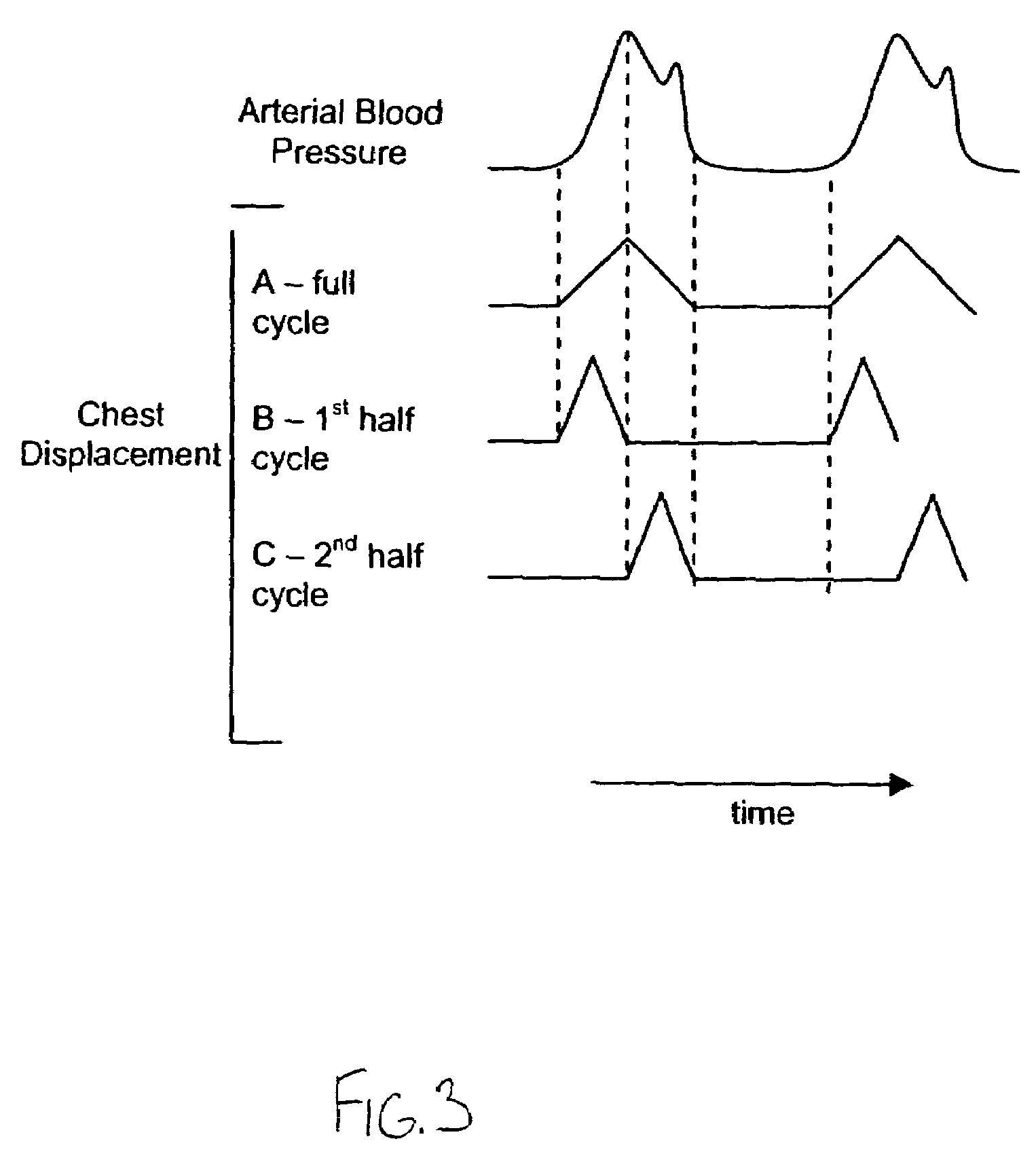
[0010]The compressive force may be applied over a wide and potentially variable range of time intervals. For example, the compressive force may be applied for only a certain portion of the contraction or ejection phase, such as at the beginning, middle or end. As another example, the compressive force may be applied during each and every sensed contraction or ejection phase, or only at certain contraction or ejection phases. In other words, the start of the compression and the duration of the compression can be adjusted to improve outcomes.
[0014]In some cases, chest compressions may be performed manually, such as using traditional CPR techniques. In such cases, an audio and / or visual signal may be produced to indicate when the ejection phase is sensed. In this way, the rescuer will be prompted as to when to apply the compressive force. The tone or volume of the synchronizing prompt may be varied so as to assist the rescuer in providing optimal CPR. In some cases, the chest, abdomen, or extremities may also be actively or passively compressed or decompressed in an alternating manner with chest compressions, and in synchronization with either cardiac ejection or filling. The starting point and length of compression or decompression can also be adjusted to improve outcomes.
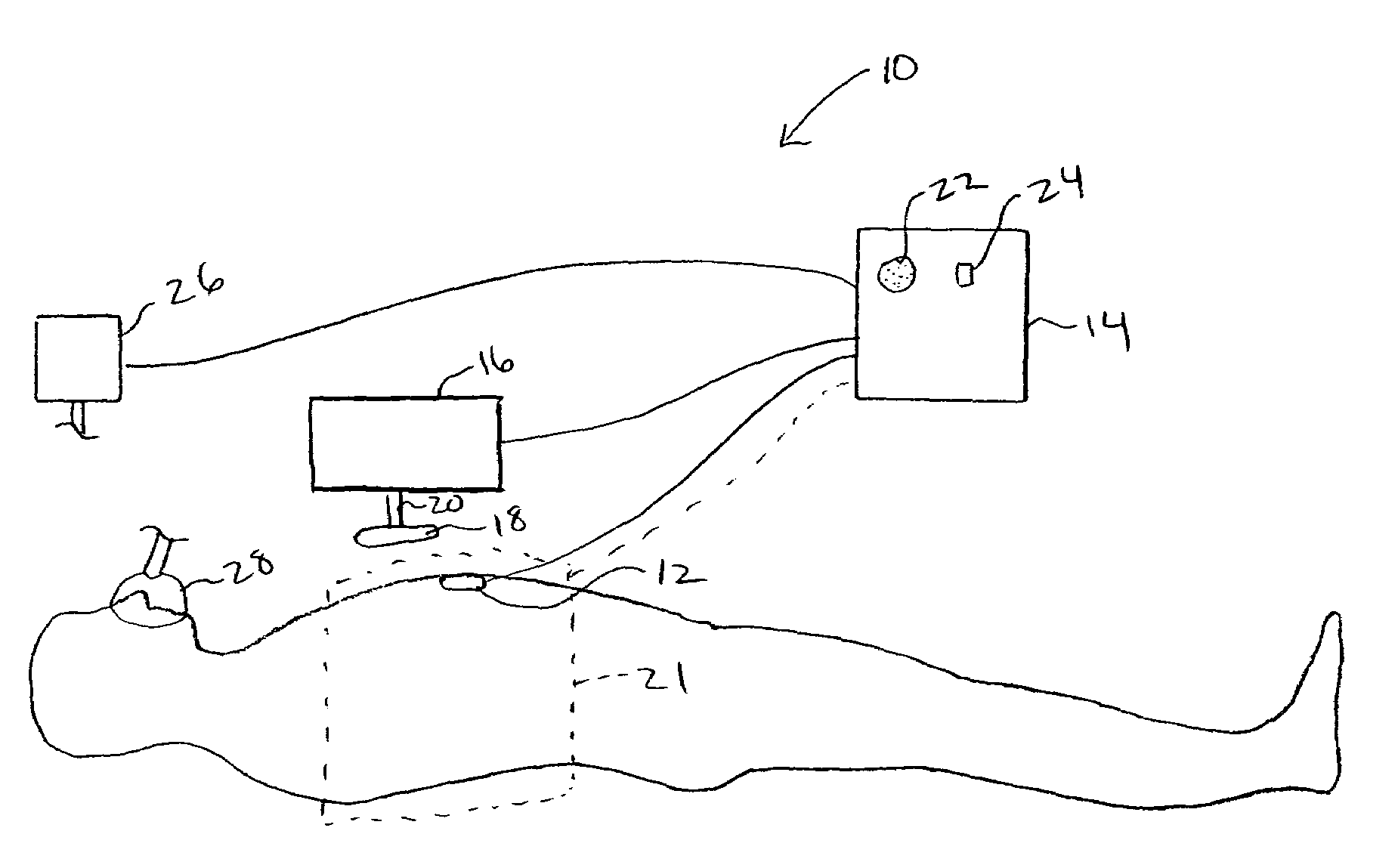
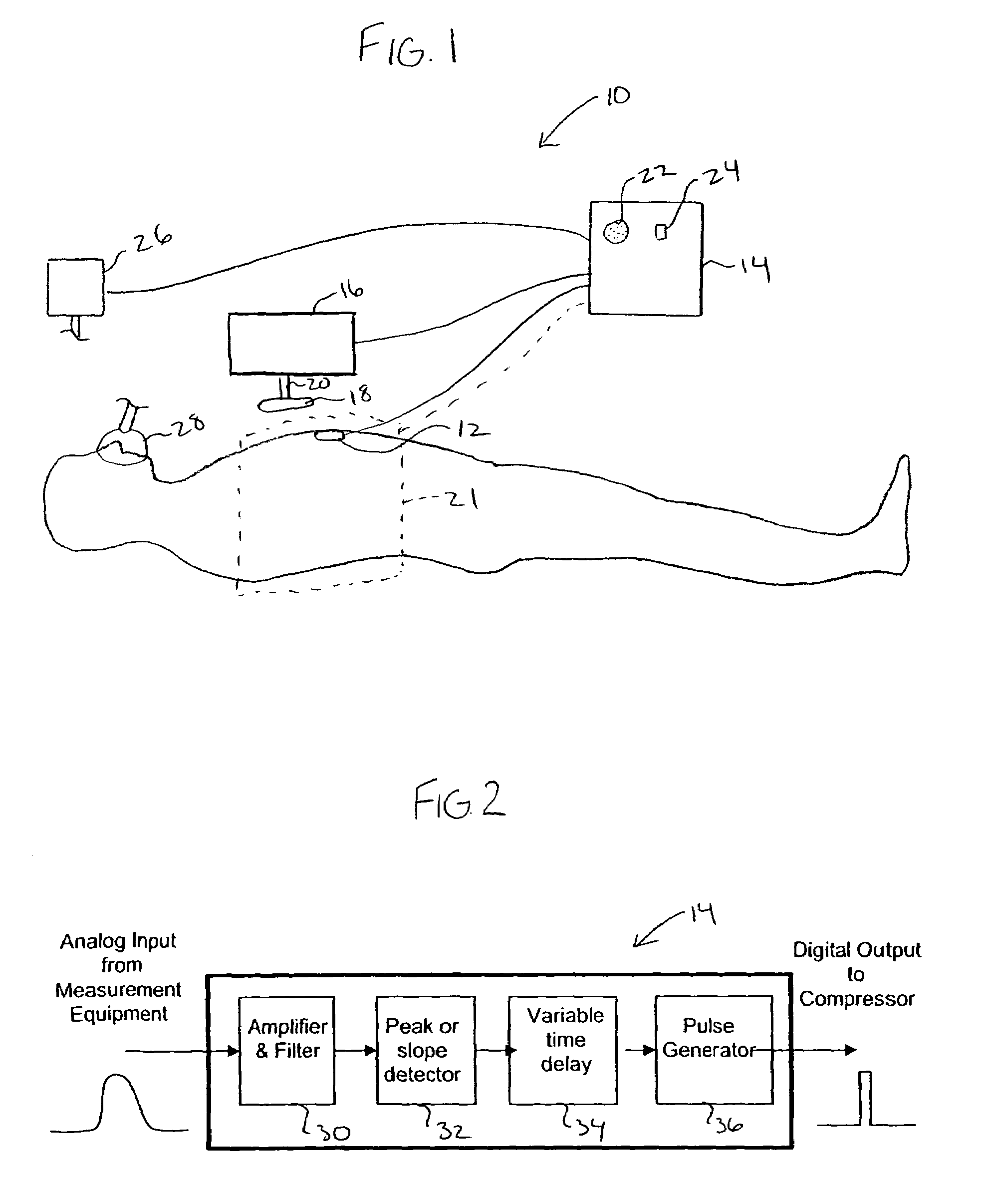
[0015]In another embodiment, the invention further provides a system for improving the cardiac output of a patient who is suffering from pulseless electrical activity or shock and yet still displays some myocardial wall motion. The system comprises a myocardial activity sensor that is adapted to sense movement of the myocardial wall and or myocardial valvular motion to determine the presence of residual left ventricular contract and relaxation, and / or pump function having an ejection phase and a filling phase. The system may also include a compression device that is configured to repeatedly apply a compressive force to the heart, either through the chest wall, intrathoracically through the pericardium, or directly to the myocardium through a endoscope and pericardial window. Further, a controller is employed to receive signals from the myocardial activity sensor and to control operation of the compression device such that the compression device repeatedly applies a compressive force to the heart such that the compressive force is applied during at least some of the ejection phases and is ceased during at least some of the relaxation phases to permit residual cardiac filling, thereby enhancing cardiac output and organ perfusion.
[0023]The vector of left ventricular blood flow ejection is generally from the point of maximal impulse in the left lateral chest between 4th and 6th intercostal spaces near the lateral clavicular line toward the medial cephalad direction. The invention could determine this vector and align the force of chest compression so as to assist ejection of blood and minimize interference with ventricular filling.

Problems solved by technology

CPR that is not synchronized with the heart's residual mechanical function may result in application of the compression phase when the left ventricle is trying to fill, resulting in significantly decreased cardiac output on the next ejection secondary to Frank-Starling Law.
Interference with ventricular filling by compression of the chest can be so deleterious that it can, in and of itself, cause complete loss of residual myocardial function resulting in true cardiac arrest.

Method used


Image

  • Non-invasive device for synchronizing chest compression and ventilation parameters to residual myocardial activity during cardiopulmonary resuscitation
  • Non-invasive device for synchronizing chest compression and ventilation parameters to residual myocardial activity during cardiopulmonary resuscitation
  • Non-invasive device for synchronizing chest compression and ventilation parameters to residual myocardial activity during cardiopulmonary resuscitation

Examples


Embodiment Construction

[0029]In a broad sense, the invention relates to techniques and devices that may be used to increase cardiac output for patients suffering from a wide variety of ailments ranging from shock to pulseless electrical activity (PEA) where the patient appears to be lifeless yet has some residual mechanical heart activity. One exemplary technique of the invention is to sense when the heart is beating and then synchronize chest compressions, or other resuscitative techniques, with movement of the myocardial wall. In this way, various techniques may be used to optimally synchronize chest compressions (or other elements of CPR) with residual left ventricular function to improve the outcome of such patients. Hence, the invention may be used to synchronize the compression force of external devices, on or around the chest, with the ejection phase of the residual left ventricular function, and the relaxation phase with residual cardiac filling. In another aspect, the invention provides various t...


PUM


Abstract

In one embodiment, a method for improving the cardiac output of a patient who is suffering from pulseless electrical activity or shock and yet still displays some myocardial wall motion comprises sensing myocardial activity to determine the presence of residual left ventricular pump function having a contraction or ejection phase and a filling or relaxation phase. In such cases, a compressive force is repeatedly applied to the chest based on the sensed myocardial activity such that the compressive force is applied during at least some of the ejection phases and is ceased during at least some of the relaxation phases to permit residual cardiac filling, thereby enhancing cardiac output and organ perfusion. Also incorporated may be a logic circuit capable of utilizing multiple sensing modalities and optimizing the synchronization pattern between multiple phasic therapeutic modalities and myocardial residual mechanical function.

Description

BACKGROUND OF THE INVENTION[0001]This invention relates generally to the field of cardiovascular medicine, and in particular to the treatment of patients suffering from a spectrum of cardiac states, ranging from shock to pulseless electrical activity, in which the patient appears to be lifeless and in cardiac arrest yet retains some mechanical activity in the myocardial wall motion.[0002]One common technique for treating persons suffering from cardiac arrest is the use of cardiopulmonary resuscitation or CPR. In this procedure, the patient's chest is repeatedly compressed, often in combination with periodic ventilations. Administration of electrical countershock and drugs intended to assist in restoration of cardiopulmonary function to chest compression and ventilation, constitutes advanced life support.[0003]For a variety of reasons, the effectiveness of CPR has been limited. Hence, devices or techniques which can improve the effectiveness of CPR are greatly needed.[0004]In additio...

Claims


Application Information

Patent Timeline
Patent Type & Authority Patents(United States)
IPC IPC(8): A61H31/00
CPCA61H31/005A61H31/006A61H31/007A61M16/00A61H2031/001A61H2201/5007Y10S601/09A61H2201/5058A61H2230/04A61M2205/332A61H2201/5048A61M16/0051A61M16/024
Inventor PARADIS, NORMAN A.
Owner ZOLL MEDICAL CORPORATION
Why Eureka
Try Eureka