
Automatic detection of regions (such as, e.g., renal regions, including, e.g., kidney regions) in dynamic imaging studies

a dynamic imaging and automatic detection technology, applied in the field of imaging systems, can solve the problems of limiting the usefulness of existing automated methods, reducing the enormous amount of data in a radionuclide dynamic study to a few, and not working well for existing automated methods

Active Publication Date: 2010-02-23
SIEMENS MEDICAL SOLUTIONS USA INC
View PDF3 Cites 1 Cited by
  • Summary
  • Abstract
  • Description
  • Claims
  • Application Information

AI Technical Summary

Benefits of technology

The present invention provides a method and system for automatically detecting kidney regions in medical images. The method involves identifying kidney regions, extracting dynamic sequences from the regions, and performing principal component analysis on the sequences separately. The system includes a camera for acquiring time-sequence images, an identification module for identifying regions of interest, an extraction module for extracting dynamic sequences, and an analysis module for performing principal component analysis. The technical effects of this invention include improved accuracy and efficiency in detecting kidney regions, which can aid in the diagnosis and treatment of kidney diseases.

Problems solved by technology

See e.g. The Use of Principal Components in the Quantitative Analysis of Gamma Camera Dynamic Studies, D. C. Barber, Phys. Med. Biol. 25 No 2 (March 1980) 283-292, incorporated by reference below (“The reduction of the enormous quantity of data in a radionuclide dynamic study to a few diagnostic parameters presents a problem.
Conventional methods of data reduction using regions-of-interest or functional images have several defects which potentially limit their usefulness.
However, existing automated methods do not work well.
In fact, this is typically performed manually, but the manual method is time consuming and requires significant expertise of the operator.
While, as described above, principal component analyses and factor analyses are well known techniques and have been implemented in some background environments, there are no existing techniques that accurately and automatically determine, e.g., the cortical region of the kidneys.

Method used


Image

  • Automatic detection of regions (such as, e.g., renal regions, including, e.g., kidney regions) in dynamic imaging studies
  • Automatic detection of regions (such as, e.g., renal regions, including, e.g., kidney regions) in dynamic imaging studies
  • Automatic detection of regions (such as, e.g., renal regions, including, e.g., kidney regions) in dynamic imaging studies

Examples


Embodiment Construction

[0041]The preferred embodiments of the present invention can significantly improve upon existing methods and / or apparatuses.

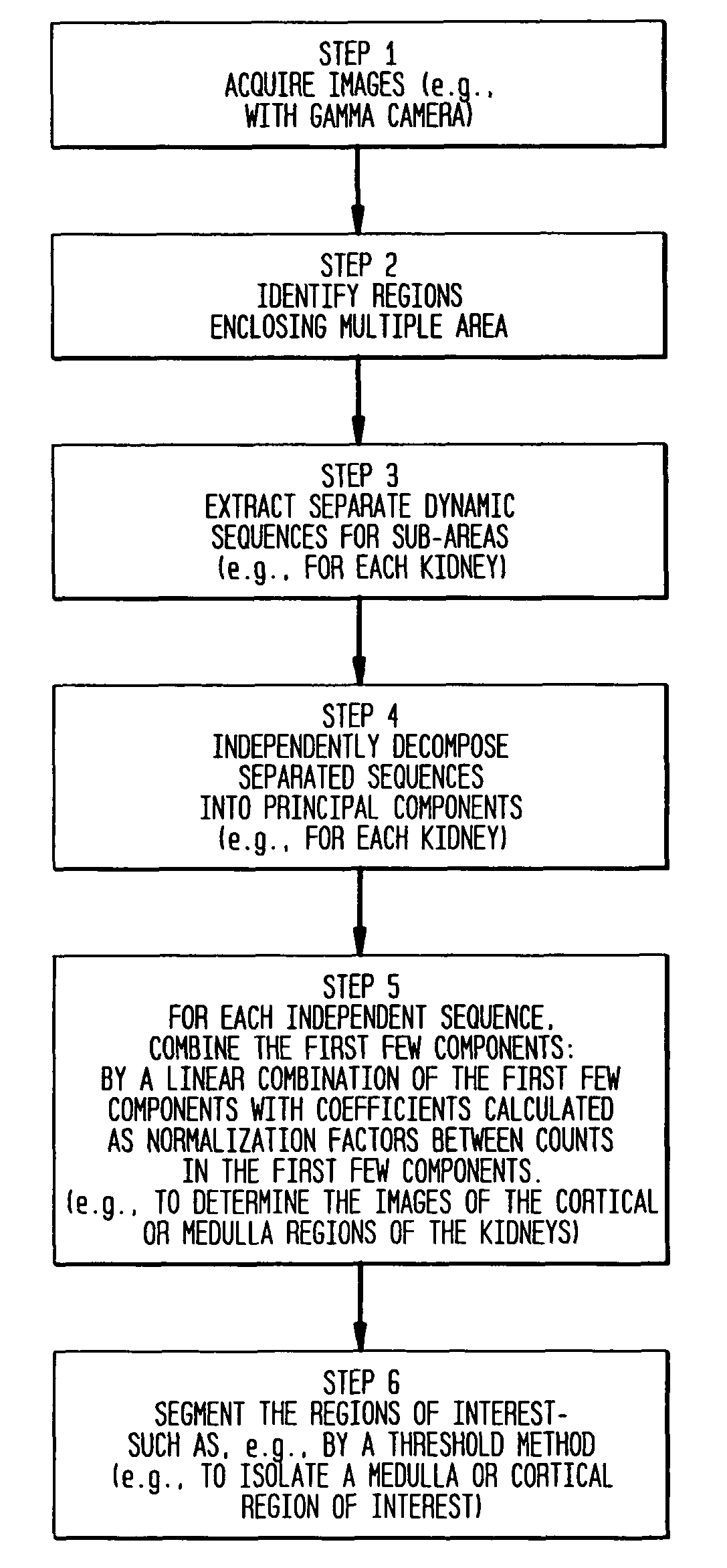
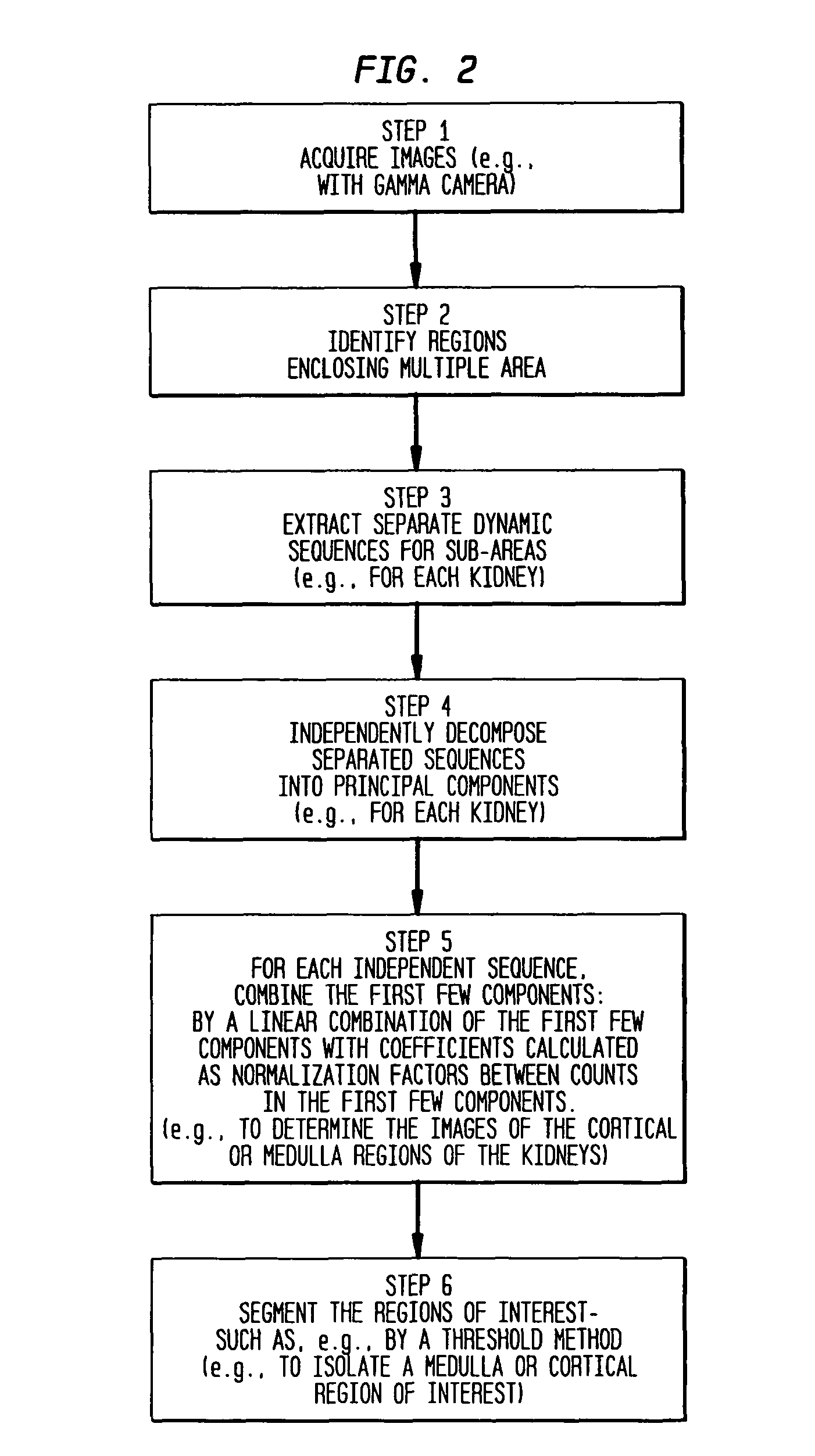
[0042]According to some preferred embodiments of the invention, a method for the automatic detection of kidney regions is performed that includes: a) identifying a first region of interest around a first kidney and a second region of interest around a second kidney; b) extracting separate dynamic sequences from the respective regions of interest; and c) performing principal component analysis on the respective dynamic sequences separately. Preferably, the performing principal component analysis includes, for each of the dynamic sequences, linearly combining the first few component images. In some embodiments, the linearly combining further includes calculating coefficients for the principal component analysis as normalization factors between counts in the first few component images. Preferably, the linearly combining further includes calculating coefficients ba...


PUM


Abstract

In some preferred embodiments, a system for the automatic identification of cortical and / or medulla regions of the kidneys in renal dynamic studies is provided that includes: a computer module configured to perform principal component analysis on a dynamic sequence corresponding to only one of the kidneys based on a linear combination of the first few component images with coefficients for the principal component analysis as normalization factors.

Description

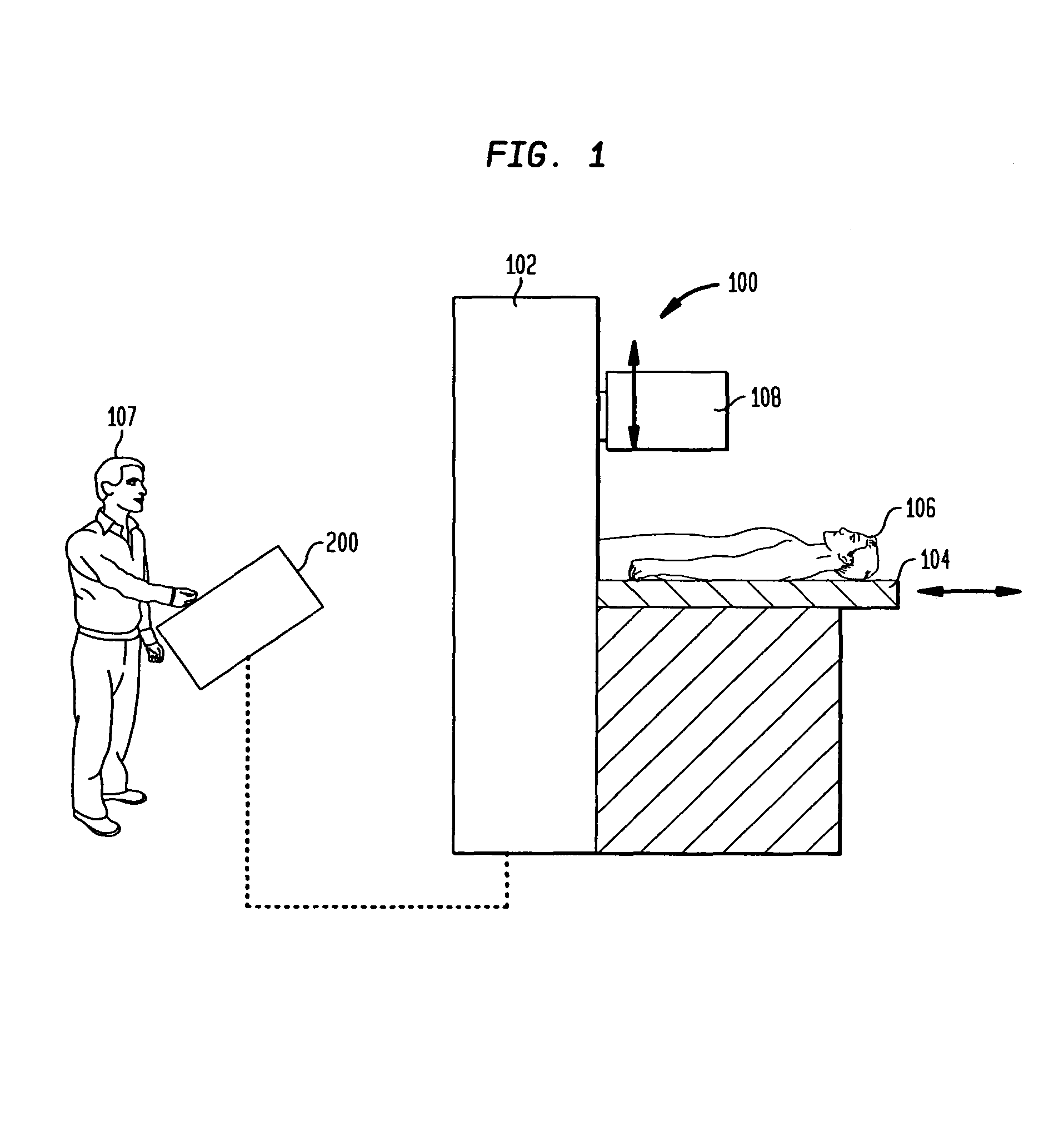
BACKGROUND[0001]1. Field of the Invention[0002]The present invention relates generally to imaging systems and, more particularly, to systems and methods for detecting regions, such as, e.g., kidney regions in renal dynamic studies.[0003]2. Discussion of the Background[0004]A variety of medical imaging systems are known. Some illustrative imaging systems include nuclear medical imaging systems (e.g., gamma cameras), computed tomography (CT or CAT) systems, magnetic resonance imaging (MRI) systems, positron-emission tomography (PET) systems, ultrasound systems and / or the like.[0005]With respect to nuclear medical imaging systems, nuclear medicine is a unique medical specialty wherein radiation (e.g., gamma radiation) is used to acquire images that show, e.g., the function and / or anatomy of organs, bones and / or tissues of the body. Typically, radioactive compounds, called radiopharmaceuticals or tracers, are introduced into the body, either by injection or ingestion, and are attracted ...

Claims


Application Information

Patent Timeline
Patent Type & Authority Patents(United States)
IPC IPC(8): G06K9/00G06V10/25
CPCG06K9/3233G06V10/25
Inventor BATTLE, XAVIER
Owner SIEMENS MEDICAL SOLUTIONS USA INC
Learn More