Nanofibrillar structure and applications including cell and tissue culture

a technology of nanofibers and nanofibers, applied in the field of nanofibers for cell culture and tissue engineering, can solve the problems of multiple layers of fine fibers, lack of an environment for living cells growth, and limited efforts

Active Publication Date: 2010-04-27
BOARD OF TRUSTEES OPERATING MICHIGAN STATE UNIV
View PDF22 Cites 69 Cited by
  • Summary
  • Abstract
  • Description
  • Claims
  • Application Information

AI Technical Summary

Benefits of technology

[0007]A nanofibrillar structure can be manufactured from a nanofiber material that provides repeatable fiber and matrix dimensions, ease of use, uniformity, cell response, quality control, and flexibility. The nanotopography, the topography of the nanofiber network of the nanofibrillar structure and the arrangement of the nanofibers of the nanofiber network in space is engineered to provide an in vitro biomimetic substratum that is more tissue-compatible for the promotion of homotypic or heterotypic cell growth and / or cell differentiation in single layer or multi-layered cell culture.

Problems solved by technology

These efforts have been limited by factors that include the use of cell lines that have been continuously grown on and selected for their ability to proliferate on planar culture surfaces that lack the spatial cues and chemical triggers and modulators present in tissue in vivo.
A media comprising multiple layers of fine fibers separated by coarse fiber supports, such as the filter media disclosed in U.S. Pat. No. 5,672,399, does not provide an environment for growth of living cells.

Method used


Image

  • Nanofibrillar structure and applications including cell and tissue culture
  • Nanofibrillar structure and applications including cell and tissue culture
  • Nanofibrillar structure and applications including cell and tissue culture

Examples


example 1

Electrospinning a Polymer Solution Comprising a Lipid Produces an Enhanced Population of Thin Fibers

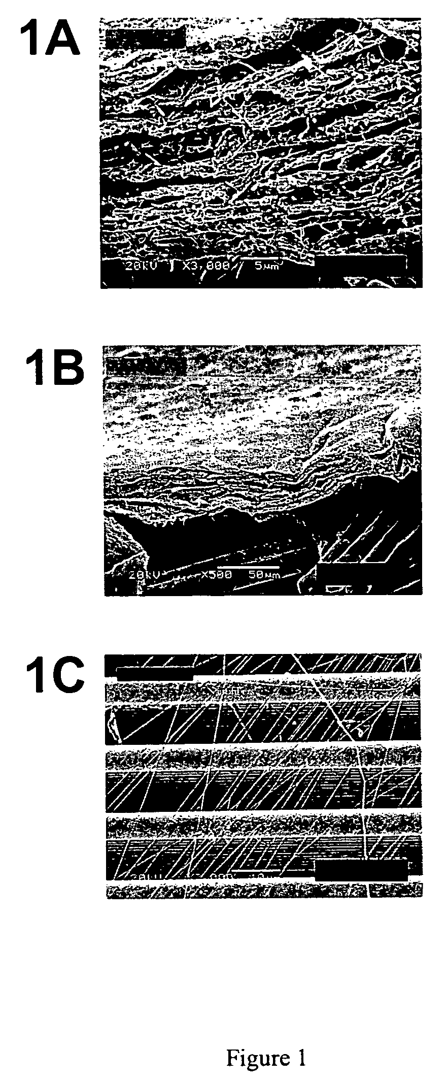
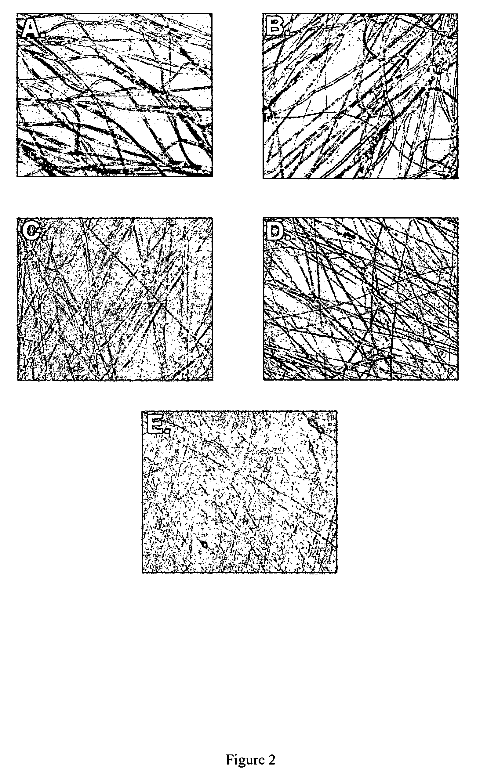
[0148]To visualize the changes in fiber diameter associated with the addition of a lipid to a polymer solution using an optical microscope, fibers were electrospun to obtain microfibers. Microfibers were electrospun from a solution comprising 15% poly(ε-caprolactone) (w / w) in chloroform supplemented with (Dow Tone Polymers, Midland, Mich.) 0, 0.25, 0.5, 1.0 and 1-% respectively of cholesterol (w / w) (Sigma, St. Louis, Mo.). The fibers were electrospun using a capillary needle system. An Eppendorf micropipette tip (yellow) was fitted to a 5 cc syringe. The polymer solution was poured into the syringe and a positive electrode connected to a Nanosecond Optical Pulse Radiator Model NR-1 (Optitron, Inc., Torrance, Calif.) was inserted into the solution. The electrospinning voltage was 18,000 volts. The fibers were electrospun onto a grounded metal plate target spinning in a plane perpendicu...

example 2

Nanofibers Comprising a Lipid Induce Tight Attachment of Cells to the Nanofiber

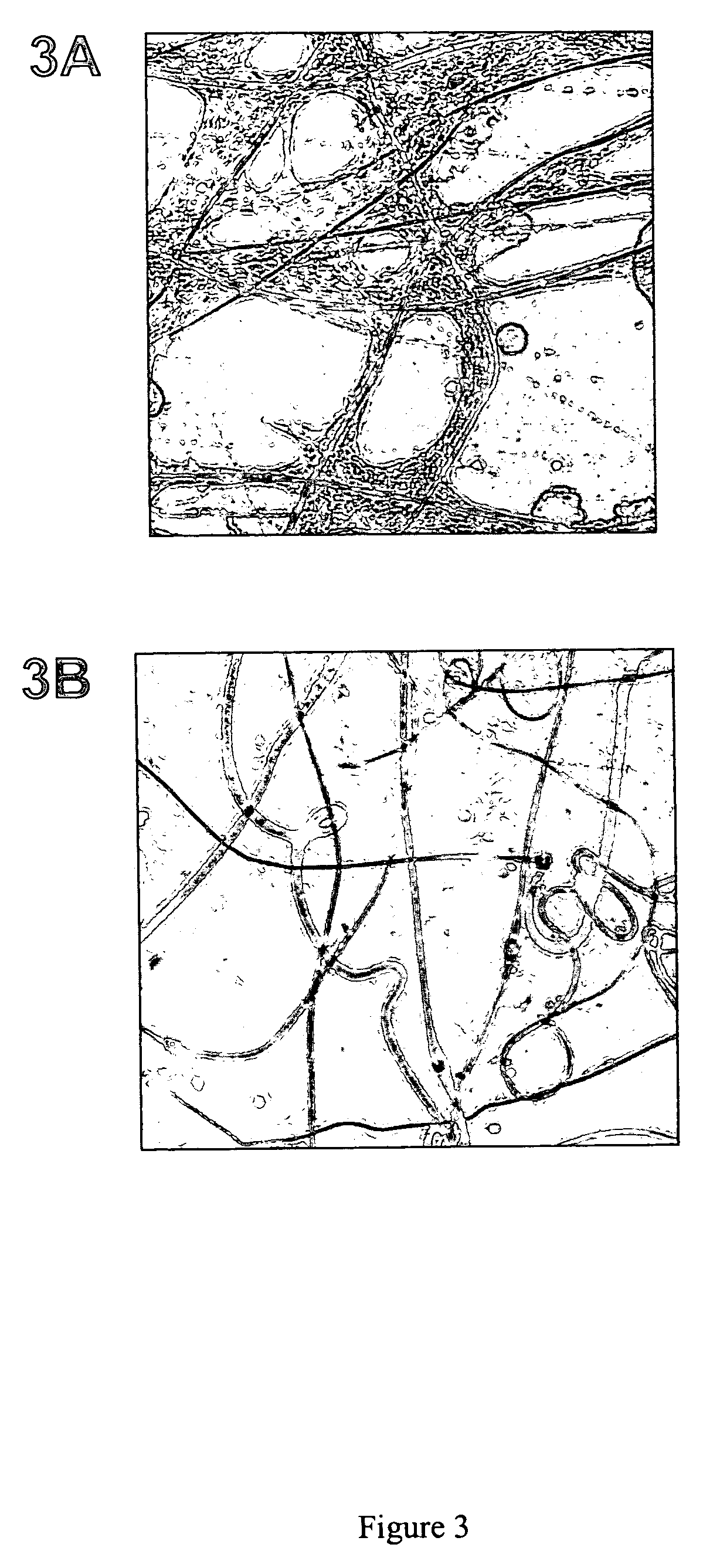
[0150]Nanofibers comprising a lipid provide a surface that promotes recruitment of cells and tight association between the cells and nanofibers. Normal kidney rat (NRK) fibroblasts were cultured on nanofibers electrospun from a solution comprising 10% poly(ε-caprolactone) (w / w) in chloroform supplemented with 0.25% sphingomyelin in Dulbecco Modified Eagle's Medium (DME) at 37° C. in 5% CO2 and visualized on a light microscope (Insight Bilateral Scanning Confocal Fluorescence Microscope (Meridian Instruments, Okemos, Mich.)) with a 20× objective. Images were captured with a CCD camera.

[0151]As shown in FIGS. 3A and B, nanofibers comprising 0.25% sphingomyelin induce a rapid recruitment of cells and their attachment to the nanofibers. FIG. 3A shows NRK fibroblasts after two days of culture on a tissue culture plate coated with nanofibers comprising 0.25% sphingomyelin. The fibroblasts are tightly attached t...

example 3

Cells Grown on Nanofiber Network have Actin Networks Similar to Cells within Tissue

[0152]The actin network of a cell has been utilized as a marker to determine which cell culture methods most closely approximate the environments within tissues (Cukierman et al., 2001, Science, 23:1708-1712; Walpita and Hay, 2002, Nature Rev. Mol. Cell. Biol., 3:137-141). When grown in two-dimensional tissue culture, fibroblasts assume a highly spread and adhering morphology in which the actin network located within the cytoplasm is organized into arrays of thick stress fibers. In contrast, fibroblasts observed in tissues are spindle-like in shape with actin organized in a cortical ring (Walpita and Hay, 2002, Nature Rev. Mol. Cell. Biol., 3:137-141).

[0153]We compared the actin network of normal rat kidney (NRK) fibroblasts grown on two-dimensional and three-dimensional surfaces. Fibroblasts were grown on polyamide nanofiber network, glass, and glass coated with polylysine. The polyamide nanofibers w...


PUM

PropertyMeasurementUnit
diameteraaaaaaaaaa
diameteraaaaaaaaaa
thicknessaaaaaaaaaa

Abstract

A nanofibrillar structure for cell culture and tissue engineering is disclosed. The nanofibrillar structure can be used in a variety of applications including methods for proliferating and / or differentiating cells and manufacturing a tissue. Also disclosed is an improved nanofiber comprising a lipid, lipophilic molecule, or chemically modified surface. The nanofibers can be used in a variety of applications including the formation of nanofibrillar structures for cell culture and tissue engineering.

Description

FIELD OF THE INVENTION[0001]The invention relates to a nanofibrillar structure for cell culture and tissue engineering and methods for proliferating and / or differentiating cells and manufacturing a tissue. Another aspect of the invention relates to a growth media for cell culture comprising a matrix of nanofibers. Another aspect of the invention relates to an improved nanofiber comprising a lipid, lipophilic molecule, or chemically modified surface. The improved nanofiber is useful in a variety of applications. In one application, a nanofibrillar structure for cell culture and tissue engineering may be prepared using the improved nanofiber. In another application, a media for cell culture may be prepared using the improved nanofiber.BACKGROUND OF THE INVENTION[0002]Cell proliferation and differentiation in vivo is regulated by unique spatial interactions between cells. Spatial cues in conjunction with the topologically distinct location of specific attachment molecules, and the rele...

Claims


Application Information

Patent Timeline
Patent Type & Authority Patents(United States)
IPC IPC(8): C12N5/00A61K49/00C08G63/06C12N5/07C12N5/071C12N5/077D01F1/02D01F6/60D01F6/62D01F11/14D04H1/4334D04H1/435D04H1/4382D04H1/728
CPCB82Y5/00C12N5/0068C12M25/14D01F1/02D01F6/60D01F6/625D01F11/14D01D5/0007C08G63/06C12N2533/40B82B3/00C12N11/02C12N5/00
Inventor SCHINDLER, MELVIN S.CHUNG, HOO YOUNG
Owner BOARD OF TRUSTEES OPERATING MICHIGAN STATE UNIV
Why Eureka
Try Eureka