Living tissue ligation device
a ligation device and living tissue technology, applied in the field of living tissue ligation device, can solve the problems of inability to obtain large torque, complicated structure, and inability to engage the arrowhead hook with the coupling member by one-touch operation, and achieve the effect of improving the gripped state of living tissue and improving the ability to stop bleeding
- Summary
- Abstract
- Description
- Claims
- Application Information
AI Technical Summary
Benefits of technology
Problems solved by technology
Method used
Image
Examples
first embodiment
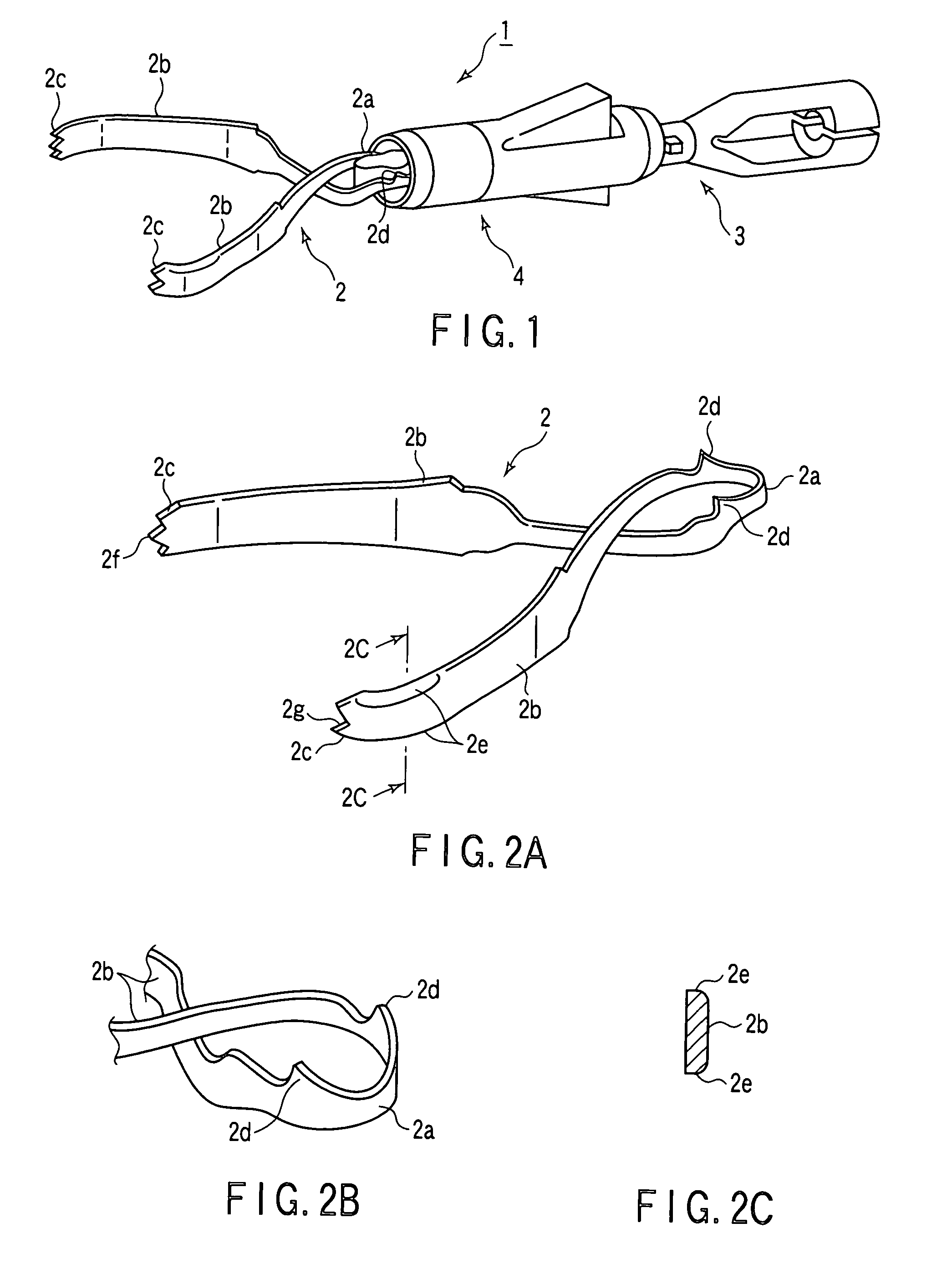
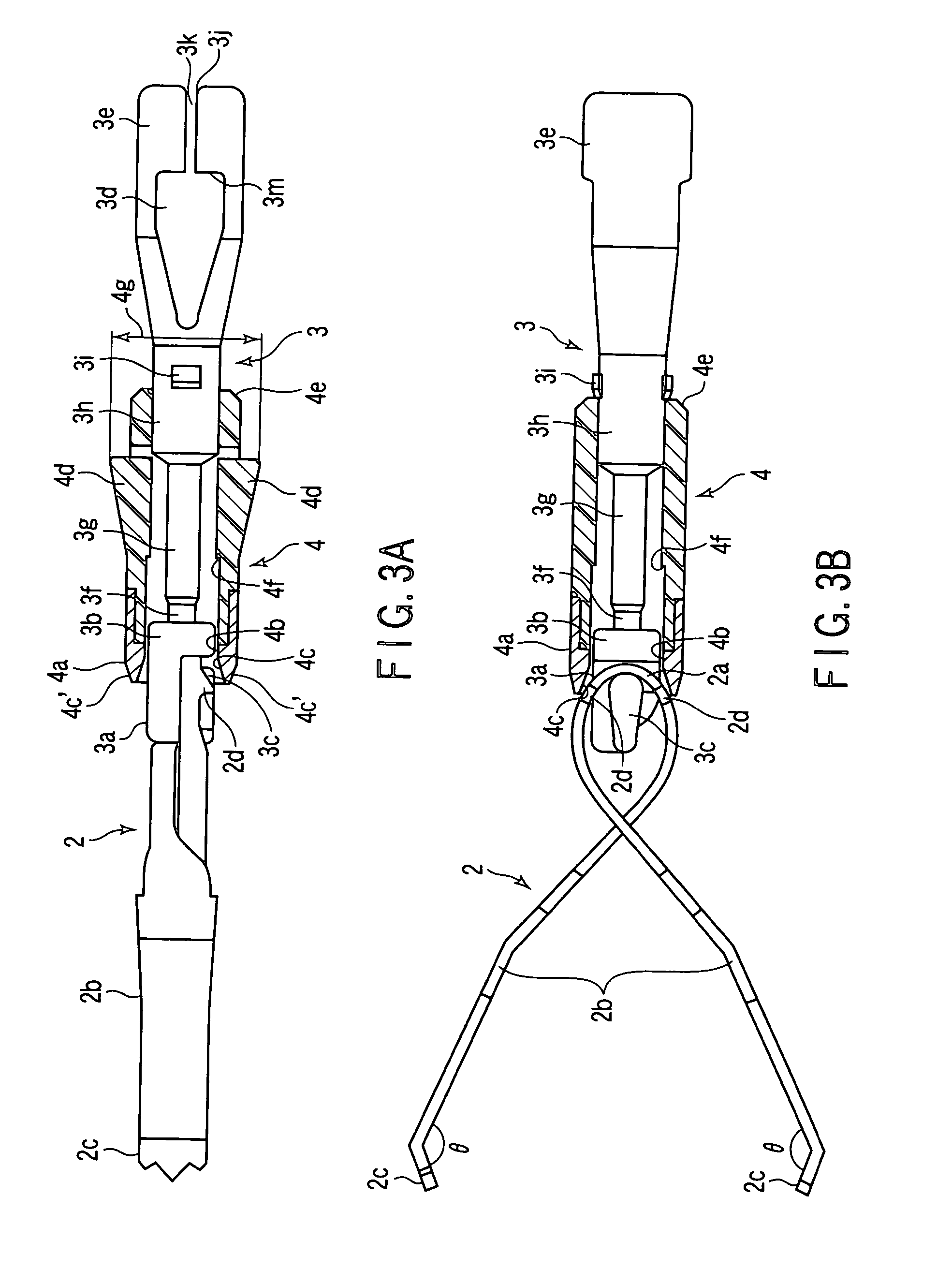
[0113]FIGS. 1 to 23 are associated with the FIG. 1 is a perspective view of a clip unit 1 of a living tissue ligation device. The clip unit 1 is comprised of a clip 2, a coupling member 3, and a press tube 4 serving as a fastening member.
[0114]As shown in FIGS. 2A to 2C, the clip 2 is formed such that after a loop portion 2a is formed by bending a metal plate member such as a leaf spring member made of stainless steel or the like at its middle portion, a pair of arms 2b having a property of spreading apart are made to intersect at a position near the loop portion 2a and extend with their distal end portions being spaced apart from each other. Tissue gripping portions 2c are formed on the distal end portions of the arms so as to face each other.
[0115]That portion of the metal plate member which corresponds to the intersecting portion of the arms 2b of the clip 2 is formed to have a width smaller than that of the distal end portions, thereby preventing the intersecting portion from b...
third embodiment
[0193]In the third embodiment, the flat surface portions 3j are formed on the shaft portion of the coupling member 3 to allow the press tube 4 to directly engage with the coupling member 3 so as to prevent the coupling member 3 from accidentally moving in the press tube 4 in the axial direction. However, the shaft portion of the coupling member 3 may be formed into a polygonal shape or elliptic shape. That is, the shape of the shaft portion is not specifically limited.
[0194]FIGS. 33A and 33B show a modification to the press tube 4. A pair of key grooves 4j are formed in that portion of the inner circumferential surface of the press tube 4 which is located on the operator side in correspondence with the pair of lock projections 3i provided on the coupling member 3 so as to be spaced apart from each other by 180°. According to this modification, at the time of assembly of the clip unit 1, the lock projections 3i engage with the key grooves 4j to restrict the coupling member 3 from mov...
seventh embodiment
[0237]FIG. 47 shows the 10th embodiment, which has the same object as that of the Referring to FIG. 47, the proximal end portion of a channel 131 incorporated in the insertion portion of an endoscope 130 is connected to a branch tube 133 inside an operating portion body 132. One tube of the branch tube 133 communicates with a push button 134 of the operating portion body 132, and the other tube communicates with a forceps opening 135. A connection tube 136 between a branch portion 133a of the branch tube 133 and the forceps opening 135 is formed to have a large inner diameter.
[0238]A large-diameter introduction tube 138 which can be inserted into the connection tube 136 is connected to the proximal end portion of the introduction tube 5 through a connection tube 137, thereby connecting the large-diameter introduction tube 138 to the operating portion body 132.
[0239]According to this embodiment, when the operating portion body 132 is rotated, the rotation can be transferred to the i...
PUM
 Abstract
Description
Claims
Application Information
 - R&D
- Intellectual Property
- Life Sciences
- Materials
- Tech Scout
- Unparalleled Data Quality
- Higher Quality Content
- 60% Fewer Hallucinations
Browse by: Latest US Patents, China's latest patents, Technical Efficacy Thesaurus, Application Domain, Technology Topic, Popular Technical Reports.
© 2025 PatSnap. All rights reserved.Legal|Privacy policy|Modern Slavery Act Transparency Statement|Sitemap|About US| Contact US: help@patsnap.com


