
Drug depot implant designs

a technology of drug depot and implant, which is applied in the direction of prosthesis, peptide/protein ingredients, antibody medical ingredients, etc., can solve the problems of limited quantity of agents, insufficient method to serve the needs of patients, and degradation of cartilage matrix, so as to facilitate implantation and retention of implants, minimize the effect of implant migration and optimal clinical efficiency

Active Publication Date: 2010-06-01
WARSAW ORTHOPEDIC INC
View PDF14 Cites 1148 Cited by
  • Summary
  • Abstract
  • Description
  • Claims
  • Application Information

AI Technical Summary

Benefits of technology

[0014]One aspect of the present invention, which provides the use of depots to deliver anti-inflammatory or anabolic compounds to intervertebral discs or articulating joints, has not been previously disclosed. Another aspect of the present invention provides specific designs and methods for insertion of drug depots into discs or joint capsules with minimal tissue disruption and minimal interference with normal joint motion. The methods, systems and reagents of the present invention prevent the depots from migrating away from the inflamed tissue and allow for more uniform distribution of the drug.
[0015]One aspect of the invention provides a method for reducing pain and / or inflammation, which comprises administering to a target site in a subject in need of treatment an effective amount of a pharmaceutical composition comprising one or more therapeutic agents, wherein the one or more therapeutic agents are administered by a drug depot implant. In one embodiment, the drug depot implant comprises a body that holds the therapeutic agent and an anchoring system that extends from the body to prevent migration of the body from the target site.
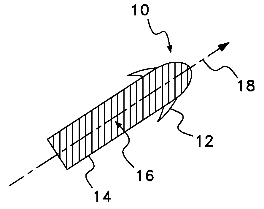
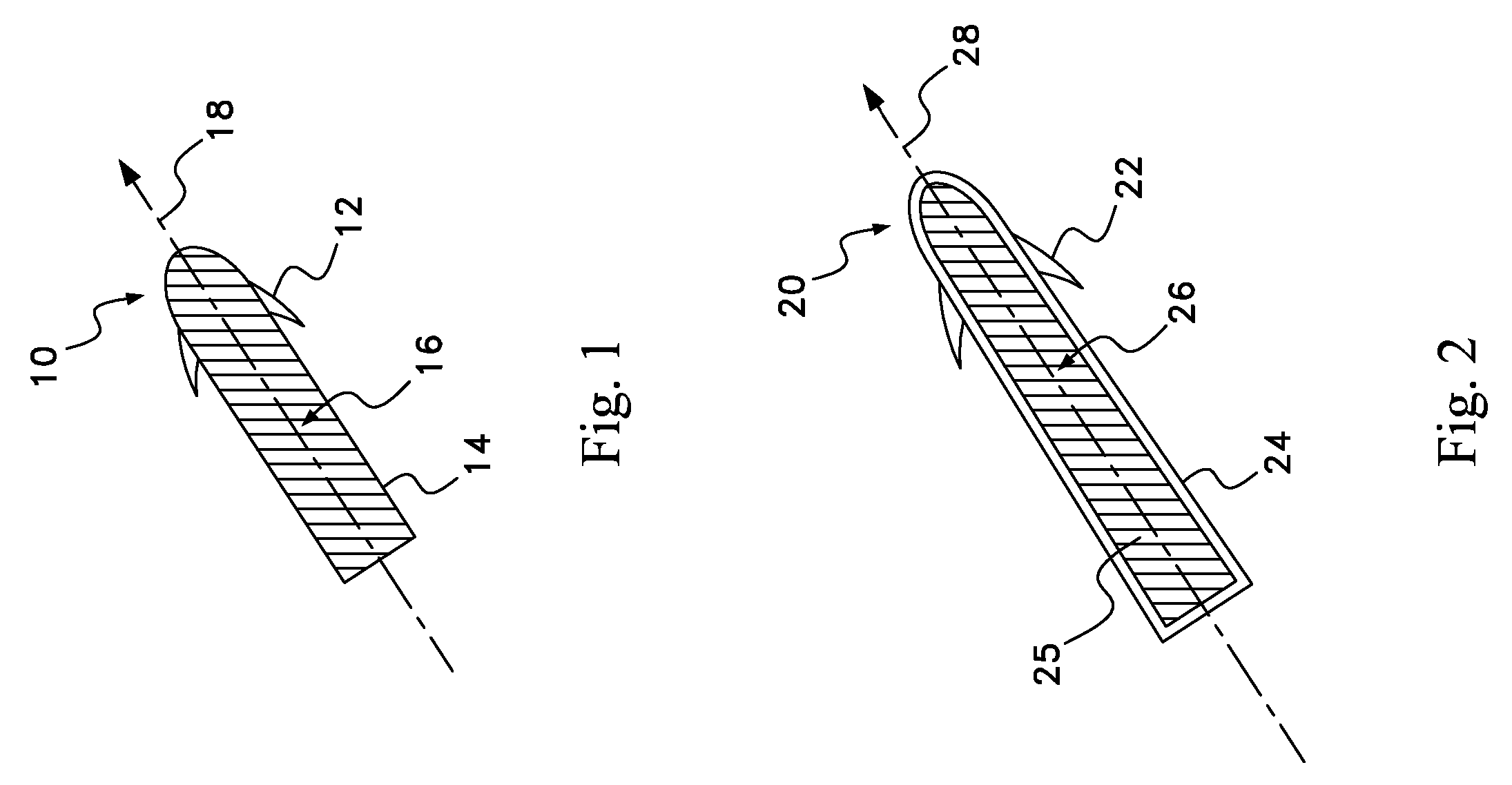
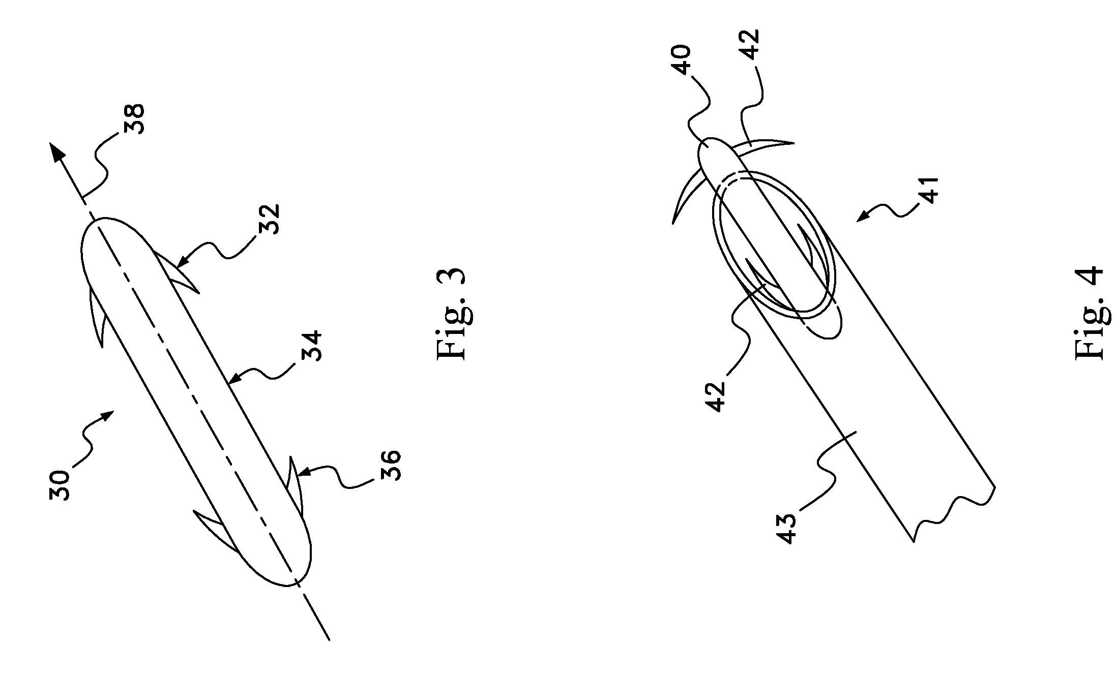
[0018]In accordance with one aspect of the present invention a drug depot implant design provides a physical feature to facilitate implantation and retention of the implant in the desired anatomical location for optimal clinical efficiency. In one embodiment of the invention the drug depot design is a rod shaped implant loaded with a therapeutic agent. One embodiment of the invention provides a rod shaped implant comprising small barbs that minimize migration of the implant in a patient's tissue once implanted.
[0019]The present invention provides for methods, systems and reagents that permit a surgeon to deliver a drug depot implant with optimal efficiency to a target site in a subject in need of treatment. In one embodiment of the invention, the implant is designed to limit “backout” or forward movement into critical tissues. In another embodiment of the invention, the implant is positioned in adjacent soft tissue to the spinal foramen space of inflamed nerve roots to alleviate sciatica and / or back pain caused by such inflammation. An alternative embodiment of the invention provides for a rod depot implant positioned into a disc space, wherein the implant is positioned in place by the barb. In this embodiment of the invention the rod depot may further comprise a built-in cap or “stop” that positions the rod in place by utilizing an adjacent tissue plane, thereby permitting the active end of the rod to protrude into an area of inflamed tissue and elute the therapeutic agent, which may be, for example, an anti-inflammatory agent. In another embodiment of the invention the drug depot implant is positioned in the knee joint, wherein the rod cap is positioned in place by the knee capsule and elutes the therapeutic agent, such as an anti-inflammatory agent, into the knee joint synovial fluid. Additional embodiments of the invention provide for positioning the drug depot implant in the shoulder, hip, other joints or spine of a patient.
[0034]One aspect of the invention provides a radiographic marker on the drug depot implant to permit a surgeon to accurately position the implant into a tissue of a patient. Examples of such radiographic markers include, but are not limited to, barium, calcium phosphate, and metal beads. Such radiographic markers will also permit the surgeon to track movement and degradation of the implant in the tissue over time. In this embodiment of the invention the surgeon may accurately position the implant in the tissue using any of the numerous diagnostic imaging procedures known to one of ordinary skill in the art. Such diagnostic imaging procedures include, for example, X-ray imaging or fluoroscopy.

Problems solved by technology

It promotes secretion of matrix metalloproteinases (MMPs), leading to cartilage matrix degradation.
TNF inhibitors currently in use are generally administered systemically via intravenous infusion and subcutaneous injection, but there are side effects of anti-TNF therapies associated with the higher doses and systemic administration that are common with these therapies.
A major disadvantage of these systemic drug delivery systems is that the anti-inflammatories are delivered in buffered solutions that have short half-lives, thereby requiring repeated administration to a patient, which can result in adverse effects due to the system of delivery of relatively high doses of the drug.
Unfortunately, it provides a limited quantity of agent that must move through the tissue to the target site.
This method is inadequate to serve the needs of patients.
In addition, injection site pain and reactions sometimes develop with anti-TNF agents.
Another drawback to this approach is that the delivery of the therapeutic agent is accomplished via the general systemic circulatory system; the therapeutic agent is not delivered locally to the affected area.
U.S. Pat. No. 6,735,475 describes a variety of implants for treating headache and / or facial pain, none of which are suitable for introducing therapeutic agents to a desired tissue of a patient.
U.S. Patent Application Publication No. 20040064193 describes a variety of implants for regeneration of tissue, none of which are suitable for introducing therapeutic agents to a desired tissue of a patient.
U.S. Patent Application Publication No. 20050074481 describes a variety of implants for facilitating the healing of voids in bone, cartilage and soft tissue, none of which are suitable for introducing therapeutic agents to a desired tissue of a patient.
One drawback of these implants is that they are fixedly attached to the bone itself in a synovial joint.
Another drawback of these implants is that many of the intended patients already suffer from pain and inflammation, and will be subjected to more pain upon implantation of the device into the bone and possible formation of osteophytes.

Method used


Image

  • Drug depot implant designs
  • Drug depot implant designs
  • Drug depot implant designs

Examples


Embodiment Construction

[0061]For the purposes of promoting an understanding of the principles of the invention, reference will now be made to preferred embodiments and specific language will be used to describe the same. It will nevertheless be understood that no limitation of the scope of the invention is thereby intended, such alterations and further modifications of the invention, and such further applications of the principles of the invention as illustrated herein, being contemplated as would normally occur to one skilled in the art to which the invention relates.

[0062]The present invention provides methods, systems and reagents for facilitating implantation of a drug depot implant into a subject comprising implanting an implant depot loaded with a therapeutic agent. The drug depot implants of the present invention may be designed for placement and location within or near the synovial joint, the spinal disc space, the spinal canal or the surrounding soft tissue.

[0063]The present invention also provid...


PUM

PropertyMeasurementUnit
timeaaaaaaaaaa
distanceaaaaaaaaaa
sizeaaaaaaaaaa

Abstract

The present invention relates to novel drug depot implant designs for optimal delivery of therapeutic agents to subjects. The invention provides a method for alleviating pain associated with neuromuscular or skeletal injury or inflammation by targeted delivery of one or more therapeutic agents to inhibit the inflammatory response which ultimately causes acute or chronic pain. Controlled and directed delivery can be provided by drug depot implants, comprising therapeutic agents, specifically designed to deliver the therapeutic agent to the desired location by facilitating their implantation, minimizing their migration from the desired tissue location, and without disrupting normal joint and soft tissue movement.

Description

CROSS REFERENCE TO RELATED APPLICATIONS[0001]This application is a continuation-in-part patent application of U.S. patent application Ser. No. 11 / 403,733, filed on Apr. 13, 2006, which is incorporated herein by reference.FIELD OF THE INVENTION[0002]The present invention broadly concerns drug depot implant designs for optimal delivery of therapeutic agents to subjects. In specific applications of the invention the compositions include novel drug depot implant designs for optimal delivery of anti-inflammatory agents and / or growth factors to inhibit or eliminate the inflammatory response that may result in acute or chronic pain and further tissue damage after disease or injury.BACKGROUND OF THE INVENTION[0003]Current approaches for treating pain and / or inflammation involve systemic delivery of therapeutic agents. Anti-inflammatory agents target tumor necrosis factor alpha (TNF-a) which appears early in the inflammatory cascade following infection or injury. It is produced by monocytes,...

Claims


Application Information

Patent Timeline
Patent Type & Authority Patents(United States)
IPC IPC(8): A61K38/16A61F2/00A61K38/17A61K38/18A61K38/19A61K38/22A61K39/00
CPCA61K9/0024A61K9/0097A61K9/0085A61P29/00
Inventor MCKAY, WILLIAM F.
Owner WARSAW ORTHOPEDIC INC
Learn More