Method and apparatus for detecting blood vessel boundaries using multi-scale mean-shift ray propagation

a mean-shift ray and blood vessel technology, applied in the field of medical diagnosis, can solve the problems of incorrect diagnosis of patients, inaccurate diagnosis of patients, and difficult task of accurate and robust detection of vessel boundaries

Active Publication Date: 2010-08-10
SIEMENS MEDICAL SOLUTIONS USA INC
View PDF10 Cites 39 Cited by
  • Summary
  • Abstract
  • Description
  • Claims
  • Application Information

AI Technical Summary

Benefits of technology

[0014]The present invention is a method and system for detecting a boundary of a vessel in an image. An accurate detection of a boundary requires an accurate detection of edges related to the vessel boundary while not recognizing edges associated with other structures unrelated to the vessel boundary. Edges are detected based on the change in intensity between data points over a plurality of distances. In one embodiment, edges are detected by propagating one or more rays along the vessel. A set of edges is then selected from the detected edges. Further, incorrect edges can be eliminated from the edges. Each edge in the selected set of edges can be selected based on its strength.

Problems solved by technology

Accurate and robust detection of vessel boundaries is traditionally a challenging task.
If the vessel boundary detection algorithm is inaccurate (even in a small number of cases), a medical professional (e.g., a radiologist) relying on the computer's output may, in turn, incorrectly diagnose the patient.
There are many reasons why accurate and robust detection of vessel boundaries is a challenging task.
First, the presence of significant noise levels in computed tomography (CT) and magnetic resonance (MR) images often forms strong edges (i.e., changes in intensity between data points) inside vessels.
Second, the size of a vessel can vary from one vessel location to another, resulting in additional edges.
Fourth, the presence of vascular pathologies, e.g., calcified plaques, often makes the shape of a vessel cross-sectional boundary locally deviate from a circular shape.
These edges can result in inaccuracy when an algorithm is used to determine the boundaries of the vessel 104.
This is an inaccurate process because the estimation of the boundary can vary widely from the actual boundary.
An inaccurate selection of one or more parameters may result in the tube expanding beyond the actual vessel boundary.
Thus, the snake model does not always provide accurate results.
However, the use of gradient strength by itself is often not enough for robust segmentation.
It is often difficult, however, to estimate parameters such as spatial, range kernel filter sizes, and / or the amount of smoothness constraints for the robust segmentation of vessels.
In particular, the use of a single spatial scale and curvature based smoothness constraints are typically not enough for accurate results when vessels are not isolated very well.

Method used


Image

  • Method and apparatus for detecting blood vessel boundaries using multi-scale mean-shift ray propagation
  • Method and apparatus for detecting blood vessel boundaries using multi-scale mean-shift ray propagation
  • Method and apparatus for detecting blood vessel boundaries using multi-scale mean-shift ray propagation

Examples


Embodiment Construction

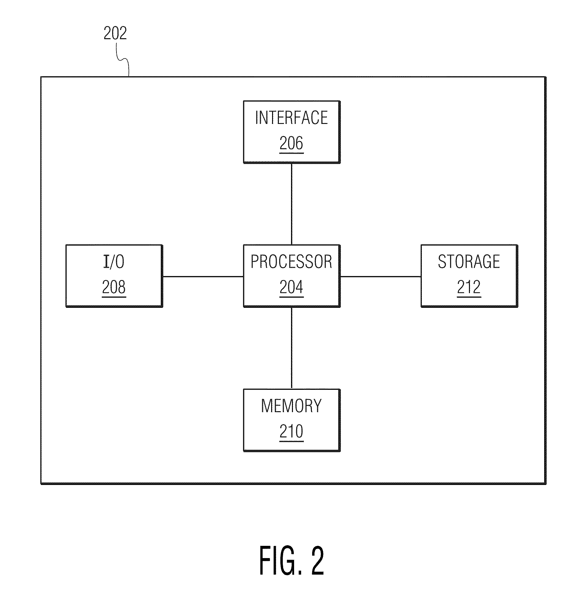
[0029]The following description describes the present invention in terms of the processing steps required to implement an embodiment of the invention. These steps may be performed by an appropriately programmed computer, the configuration of which is well known in the art. An appropriate computer may be implemented, for example, using well known computer processors, memory units, storage devices, computer software, and other components. A high level block diagram of such a computer is shown in FIG. 2. Computer 202 contains a processor 204 which controls the overall operation of computer 202 by executing computer program instructions which define such operation. The computer program instructions may be stored in a storage device 212 (e.g., magnetic disk) and loaded into memory 210 when execution of the computer program instructions is desired. Computer 202 also includes one or more interfaces 206 for communicating with other devices (e.g., locally or via a network). Computer 202 also...


PUM


Abstract

Disclosed is a method and system for detecting a boundary of a vessel in an image. Edges in the image are detected. Edge detection is based on the change in intensity over some distance while varying the scale of the distance. A set of edges is then selected from the detected edges. An initial vessel boundary is determined based on the selected set, and a shape descriptor (e.g., one or more elliptical shape descriptors) is applied to the initial vessel boundary to determine a final vessel boundary.

Description

[0001]This application claims the benefit of U.S. Provisional Application No. 60 / 672,634 filed Apr. 19, 2005, which is incorporated herein by reference.BACKGROUND OF THE INVENTION[0002]The present invention relates generally to medical diagnostics, and more particularly to the determination of vessel boundaries in a medical image.[0003]To diagnose a problem of a patient, medical professionals often have to examine the patient's vessels (e.g., blood vessels). To illuminate a vessel so that the medical professional can examine the vessel, a patient consumes (e.g., drinks) a contrast-enhancing agent. The contrast-enhancing agent brightens one or more vessels relative to the surrounding area.[0004]The main goal of the majority of contrast-enhanced (CE) magnetic resonance angiography (MRA) and computed tomography angiography (CTA) is diagnosis and qualitative or quantitative assessment of pathology in the circulatory system. Once the location of the pathology is determined, quantitative ...

Claims


Application Information

Patent Timeline
Patent Type & Authority Patents(United States)
IPC IPC(8): G06K9/00G06K9/34
CPCG06T7/0083G06T7/60G06T2207/10072G06T2207/20116G06T2207/20136G06T2207/20156G06T2207/20192G06T2207/30101G06T7/12G06T7/181
Inventor TEK, HUSEYINAYVACI, ALPERCOMANICIU, DORIN
Owner SIEMENS MEDICAL SOLUTIONS USA INC
Why Eureka
Try Eureka