Virtual microscope system for monitoring the progress of corneal ablative surgery and associated methods

a technology of corneal ablative surgery and virtual microscope, which is applied in the field of surgical methods, can solve the problems of compromising work flow and white light not providing optimal enhancement of eye parts for visualization, and achieves the effect of improving the efficiency of surgical operations and improving the quality of surgical procedures

Active Publication Date: 2010-08-31
ALCON INC
View PDF6 Cites 24 Cited by
  • Summary
  • Abstract
  • Description
  • Claims
  • Application Information

AI Technical Summary

Problems solved by technology

However, the flooding of the eye with such illumination to detect flap position, debris, and hydration can be uncomfortable for the patient, and the use of a slit lamp to detect flap replacement and general eye condition can compromise work flow.
Further, white light may not provide optimal enhancement of parts of the eye for visualization.

Method used


Image

  • Virtual microscope system for monitoring the progress of corneal ablative surgery and associated methods
  • Virtual microscope system for monitoring the progress of corneal ablative surgery and associated methods
  • Virtual microscope system for monitoring the progress of corneal ablative surgery and associated methods

Examples


Embodiment Construction

[0018]A description of the preferred embodiments of the present invention will now be presented with reference to FIGS. 1-7.

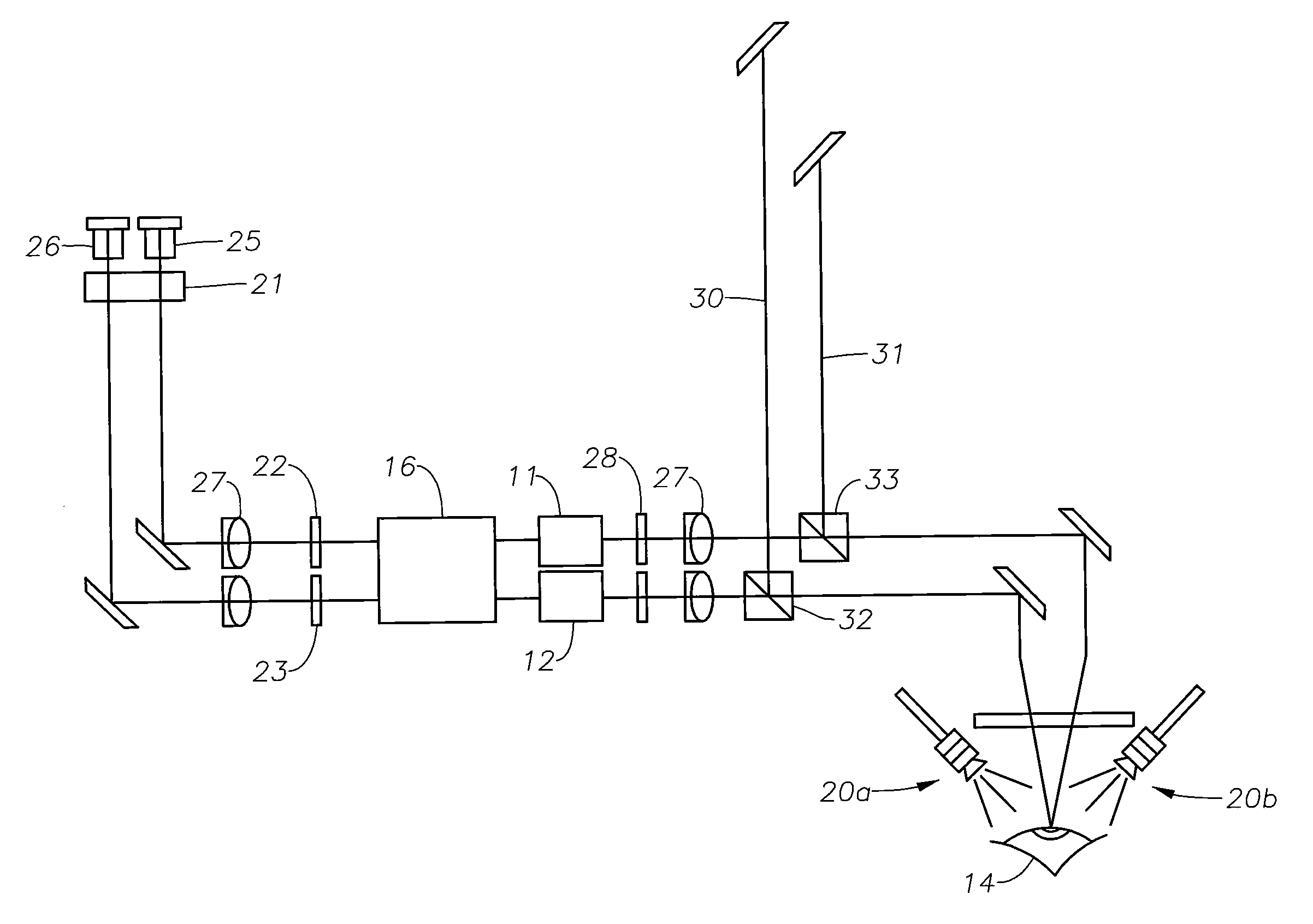
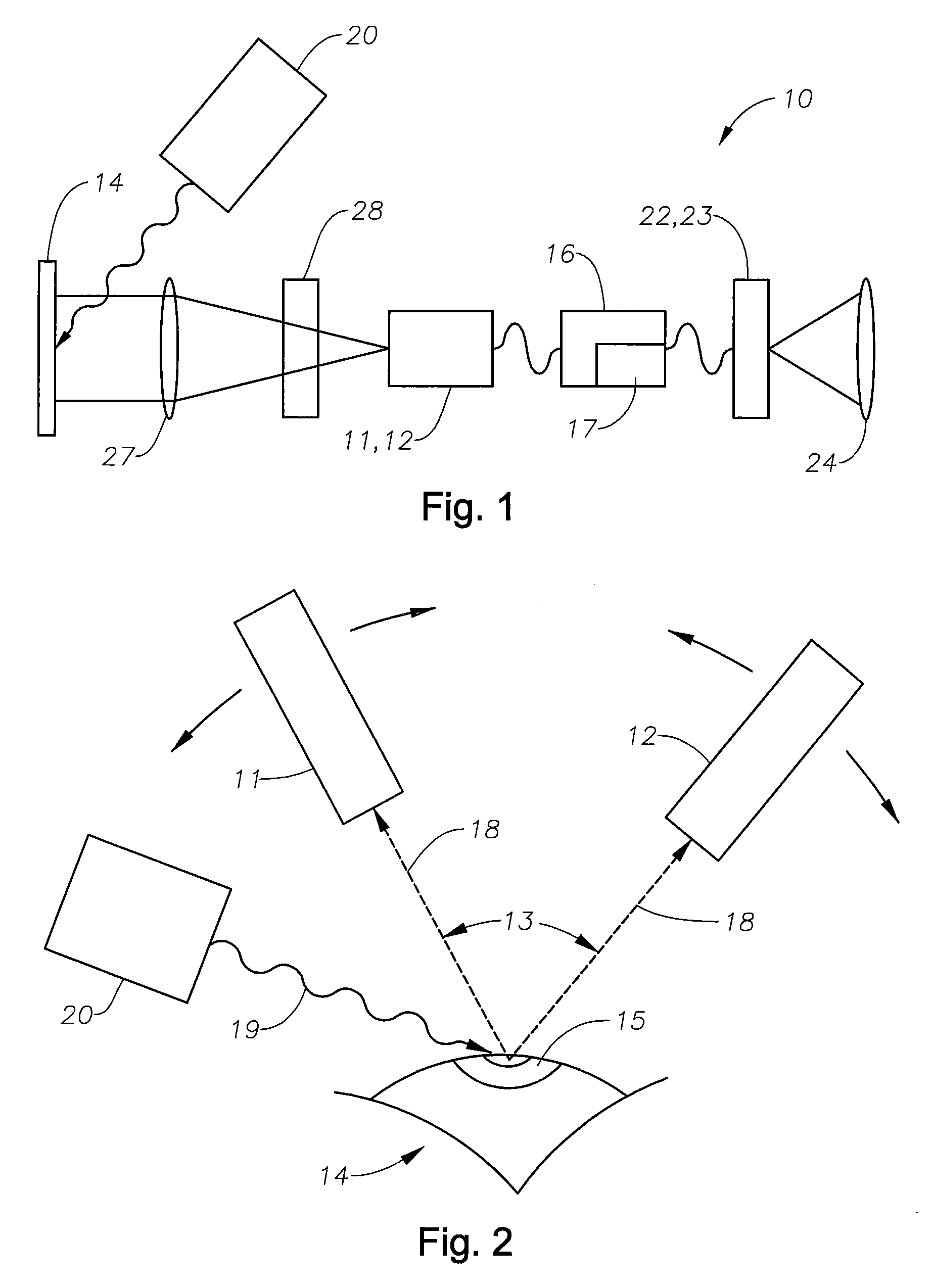
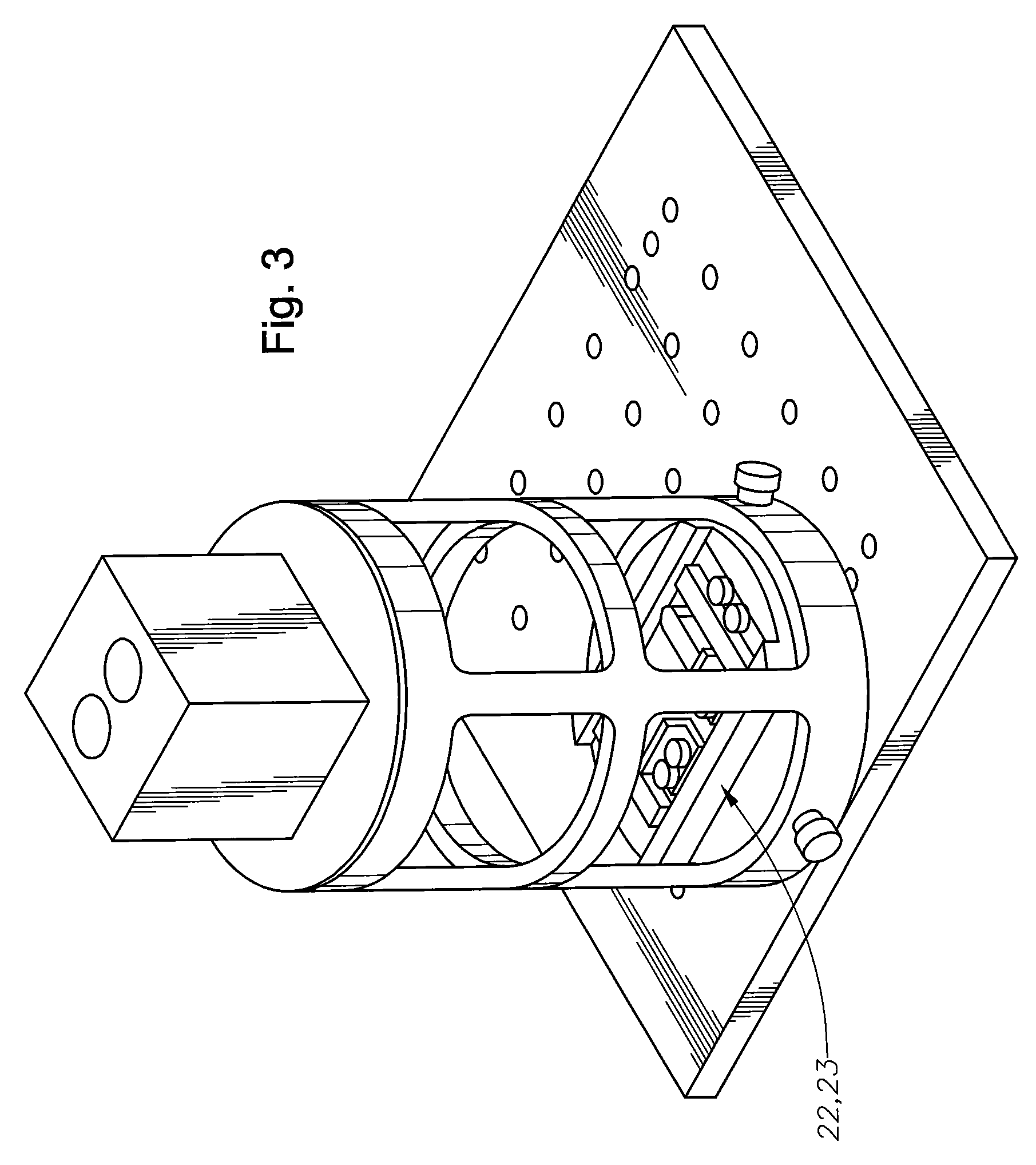
[0019]The system schematic of FIG. 1 illustrates the elements of an exemplary embodiment of a system 10 of the present invention for monitoring a process of corneal surgery by a surgeon. The system 10 comprises a first 11 and second 12 high-resolution color camera (FIG. 2) that in a particular embodiment are adjustable in angular separation 13 and can focus on a portion of an eye 14, for example, the cornea 15. An exemplary surgical procedure for which the system 10 is applicable is LASIK surgery, although this is not intended as a limitation, and is also applicable to pupilometry, where pupil dynamics can be monitored and recorded, and other eye measurements, such as corneal birefringence, and to other ophthalmic surgeries, where a surgical microscope might be useful. For this type of surgery, the system 10 can be useful for imaging the cornea 15, a flap cut i...


PUM


Abstract

A system for visualizing an eye of a patient during corneal surgery includes a processor and a first and second camera in signal communication with the processor. The cameras are positionable for focusing on a cornea positioned for surgery. A first and a second display and optics therefor are in signal communication with the processor and are positionable for viewing through a first and a second eyepiece of a stereo microscope, respectively. Software is resident on the processor for receiving a first and second corneal image from the first and second cameras, for processing the received first and second images for display, and for transmitting the processed first and second images to the first and the second displays, respectively, via the display optics. The displays can then be viewed by a surgeon through the microscope at least during the surgery.

Description

[0001]This Application Claims the Priority of U.S. Provisional Application No. 61 / 015,853 Filed Dec. 21, 2007.FIELD OF THE INVENTION[0002]The present invention generally relates to surgical methods and, in particular, to systems and methods for monitoring the progress of corneal ablative surgery.BACKGROUND[0003]The performance of LASIK (laser in situ keratomileusis) surgery is typically accompanied by the cutting of a thin flap in the cornea, which is then lifted and folded back along a hinge to expose the corneal stroma beneath. An ablating laser is used to perform refractive surgery, and the flap is replaced.[0004]Several methods have been used to avoid or detect any “wrinkles,” or striae, in the corneal flap after replacement atop the stroma. For example, the cornea can be marked prior to cutting so that the markings can be used to realign the flap. Another method employs the operating (direct-view) microscope and a diffuse, broadband, white light source to detect striae. Alterna...

Claims


Application Information

Patent Timeline
Patent Type & Authority Patents(United States)
IPC IPC(8): A61B3/14
CPCA61B3/132A61F9/008A61F2009/00844A61F2009/00872
Inventor LEBLANC, RICHARD ALAN
Owner ALCON INC
Why Eureka
Try Eureka