System and method of surgical imagining with anatomical overlay for navigation of surgical devices

a surgical device and anatomical overlay technology, applied in the field of interventional medicine, can solve the problem of difficult for physicians to become oriented in a three-dimensional setting using a single-plane x-ray image projection display

Active Publication Date: 2010-11-09
STEREOTAXIS
View PDF108 Cites 217 Cited by
  • Summary
  • Abstract
  • Description
  • Claims
  • Application Information

AI Technical Summary

Benefits of technology

[0005]According to the principles of the present invention, a system and method are provided for control of a navigation system for deploying a medical device within a subject, and for enhancement of a display image of anatomical features for viewing the current location and orientation of a medical device moving through the subject body. The display of the X-ray imaging system information is augmented in a manner such that a physician can more easily become oriented in three dimensions with the use of a single-plane X-ray display. A typical X-ray imaging system comprises a source for emitting a beam through a three dimensional space and onto a plane, where a point within a subject body in the three dimensional space is projected onto the plane. The projection of a point within the subject body onto the imaging plane can be obtained using an orthographic projection matrix derived from the point-to-image plane distance and the source-to-image plane distance. Thus, a point location within the subject body having known coordinates, properly registered to the frame of reference of the X-ray system, can be projected onto the X-ray image plane of the live X-ray image in the same manner.
[0006]In accordance with one aspect of the invention, a method of projection can be used to graphically overlay a representation of the actual medical device location and orientation onto the X-ray image. One or more desired target points within the subject can also be projected onto the X-ray image, as well as one or more reference markers on the subject to track patient movement. A graphical representation of a virtual medical device can be overlaid to show a visual reference of a predicted new location and orientation of the actual medical device that corresponds to a desired navigational configuration. A mathematical model of the medical device can be used to define the configuration of the virtual medical device, which can model the behavior of the device corresponding to a change in navigation control variables to predict deflection and rotation of the medical device. A desired direction for steering the medical device within the plane of the X-ray image can be graphically represented, and surface shapes within the subject may also be rendered and graphically represented on the X-ray image display. All the graphically overlaid information is also updated in real time as the X-ray imaging system is rotated or moved, to augment the image display and enhance visualization of the orientation of a medical device in a three dimensional space using a single-plane X-ray image displayed on the control system.
[0007]It is thus an object of the invention to provide a system and method for augmenting the displayed anatomical image of a subject with graphically overlaid objects to provide enhanced visualization of medical devices, anatomical locations, shapes, markers, and other objects and annotations in a three dimensional space for aiding in the orientation and navigation of the medical device through the subject body.

Problems solved by technology

In some cases, it may be difficult for a physician to become oriented in a three dimensional setting using a display of a single-plane X-ray image projection.

Method used


Image

  • System and method of surgical imagining with anatomical overlay for navigation of surgical devices
  • System and method of surgical imagining with anatomical overlay for navigation of surgical devices
  • System and method of surgical imagining with anatomical overlay for navigation of surgical devices

Examples


Embodiment Construction

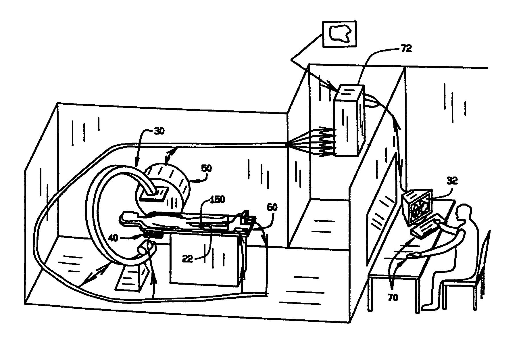
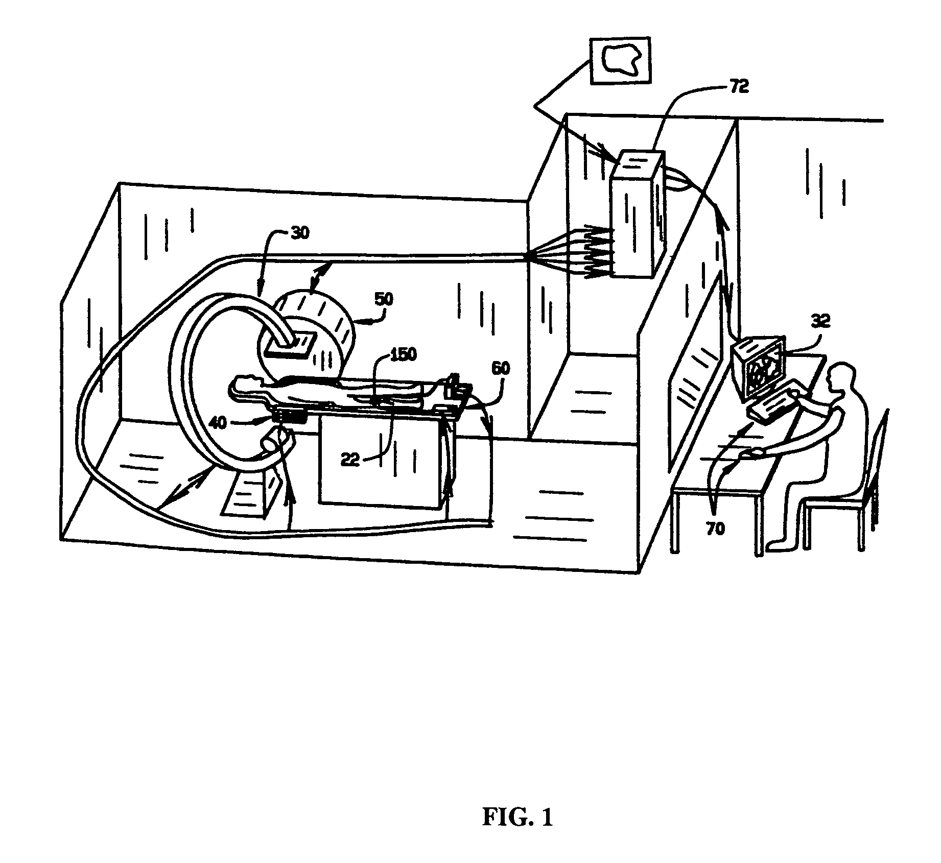
[0027]An automated system for navigating a medical device through the lumens and cavities in an operating region in a patient in accordance with the principles of this invention is indicated generally as 20 in FIG. 1. The system 20 comprises an elongate medical device 22, having a proximal end and a distal end adapted to be introduced into the operating region in a subject. The system 20 also comprises an imaging system 30 for displaying an image of the operating region on a display 32, including a representation of the distal end of the medical device 22 in the operating region.

[0028]The system also includes a navigation system for manipulating the distal end of the medical device 22. In this preferred embodiment the navigating system is a magnetic navigation system 50. Of course, the navigation system could alternatively be a piezoelectric or electrostrictive system or a mechanical control system with pull wires or servo motors, or other suitable system for orienting the distal ti...


PUM

PropertyMeasurementUnit
imaging parametersaaaaaaaaaa
imagingaaaaaaaaaa
live imaging displayaaaaaaaaaa

Abstract

A system and method are provided for control of a navigation system for deploying a medical device within a subject, and for enhancement of a display image of anatomical features for viewing the projected location and movement of medical devices, and projected locations of a variety of anatomical features and other spatial markers in the operating region. The display of the X-ray imaging system information is augmented in a manner such that a physician can more easily become oriented in three dimensions with the use of a single-plane X-ray display. The projection of points and geometrical shapes within the subject body onto a known imaging plane can be obtained using associated imaging parameters and projective geometry.

Description

FIELD OF THE INVENTION[0001]This invention relates to a system and methods for interventional medicine, and more specifically to computer assisted navigation and imaging of medical devices within a subject body.BACKGROUND OF THE INVENTION[0002]Interventional medicine is the collection of medical procedures in which access to the site of treatment is made through one of the subject's blood vessels, body cavities or lumens. For example, angioplasty of a coronary artery is most often performed using a catheter which enters the patient's arterial system through a puncture of the femoral artery in the groin area. Other interventional medical procedures include the assessment and treatment of tissues on the inner surface of the heart (endocardial surfaces) accessed via peripheral veins or arteries, treatment of vascular defects such as cerebral aneurysms, removal of embolic clots and debris from vessels, treatment of tumors via vascular access, endoscopy of the intestinal tract, etc.[0003...

Claims


Application Information

Patent Timeline
Patent Type & Authority Patents(United States)
IPC IPC(8): A61B5/05
CPCA61B5/062A61B6/12A61B6/463A61B6/547A61B19/52A61B19/5244A61B6/4441A61B19/50A61B19/56A61B2017/00243A61B2017/00703A61B2019/501A61B2019/505A61B2019/507A61B2019/5238A61B2019/5251A61B2019/5287A61B2019/5289A61B2019/5291A61B2034/101A61B90/36A61B2090/363A61B34/20A61B34/25A61B2090/364A61B2090/376A61B34/10A61B2034/105A61B2034/107A61B2034/2051A61B2090/365
Inventor VISWANATHAN, RAJU R.
Owner STEREOTAXIS
Why Eureka
Try Eureka