Efficient border extraction of image feature

a border extraction and image technology, applied in image enhancement, medical/anatomical pattern recognition, instruments, etc., can solve the problems of difficult to determine the boundary of the breast in x-ray images, time-consuming and expensive computer power requirements, and difficult to include all tissues precisely

Active Publication Date: 2011-03-29
SIEMENS COMP AIDED DIAGNOSIS
View PDF7 Cites 55 Cited by
  • Summary
  • Abstract
  • Description
  • Claims
  • Application Information

AI Technical Summary

Problems solved by technology

Since the density, size and texture of breasts are so very variable, determination of the boundary of the breast in x-ray images is not easy and current algorithms are time consuming and expensive in terms of the computer power required.
Such methods make it very difficult to precisely include all tissue, especially the peripheral skin or other border areas having low tissue density.

Method used


Image

  • Efficient border extraction of image feature
  • Efficient border extraction of image feature
  • Efficient border extraction of image feature

Examples


Embodiment Construction

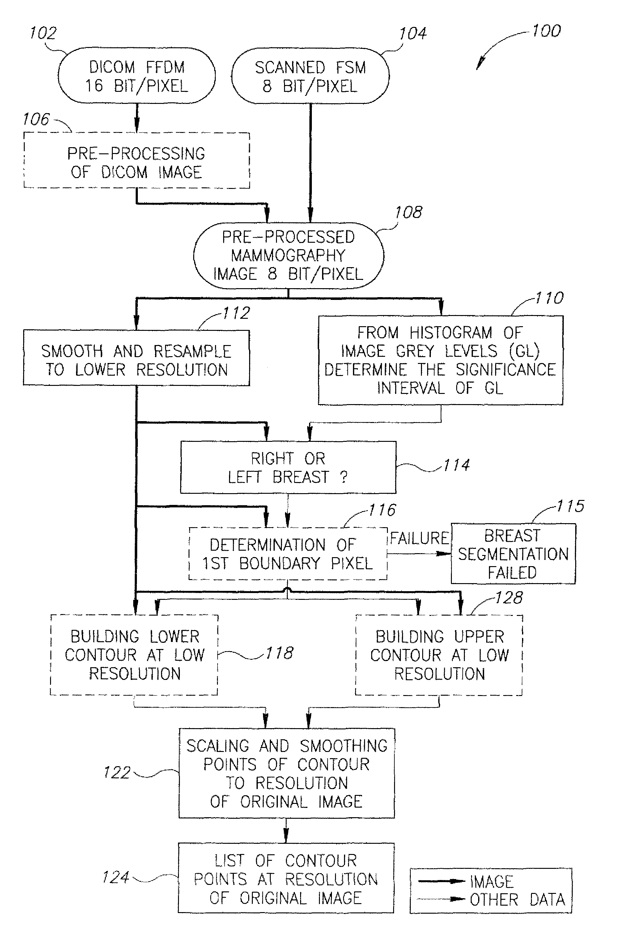
[0121]The present invention relates to determining the boundary or edge of a feature shown in an image. By way of example, the important and challenging field of breast segmentation is related to hereunder. It will be appreciated however, that the methodology disclosed herein is useful in other image analysis applications in general and medical imaging in particular.

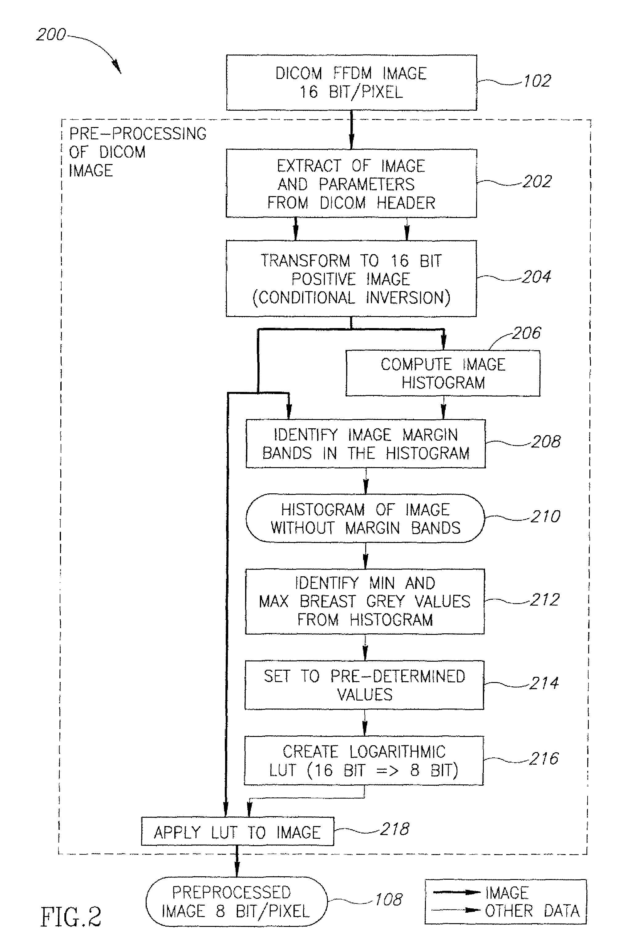
[0122]Once the breast image is obtained, using mammography techniques as known, where the image is a digital image, a pre-processing method is first applied on the original Digital Imaging and COmmunication in Medicine (DICOM) image in order to standardize the image requiring breast segmentation. This preprocessing is essentially a normalization of the image so that contrast depends more on the physiology of the breast than on the imaging parameters.

[0123]After removal of the white border strips that are sometimes evident in such images, the minimum and maximum grey values (i.e. brightness) of the image are determined an...


PUM


Abstract

Method of segmenting mammography image comprising steps of: scanning along row from outermost pixel towards the breast and comparing grayscale gradient across adjacent pixels until a significant gradient is determined; confirming that variation in gradient indicates a first true boundary pixel by ascertaining gradient across subsequent pixels is still significant;iteratively repeating steps of:(i) scanning next adjacent line in vicinity of boundary pixel, and(ii) determining the boundary pixel of adjacent line by variation in grayscale intensity gradient along row(iii) until failure to find subsequent pixel occurs and(iv) attempting scanning horizontally by repeating steps (i) and (ii), and, if attempt fails, extrapolating to edge of image or to first line, until having determined boundary around the feature to one side of first line;repeating steps (i) to (iv) on other side of line until boundary of breast on the other side of the line is determined.

Description

PRIORITY[0001]This application claims priority from U.S. Provisional Application Ser. No. 60 / 844,378 filed on Sep. 14, 2006.FIELD OF THE INVENTION[0002]The present invention is directed to image analysis, specifically to locating the boundary of a feature in an image, such as of specific tissue type in an X-ray image, particularly but not exclusively for breast segmentation of a digital mammography image.BACKGROUND OF THE INVENTION[0003]For many image analysis applications it is necessary to identify the boundary of features shown in an image. Particularly in medical imaging, such as X-ray images of lungs or breasts for diagnosing cancerous tumors and the like, it is essential to identify the boundary of the lung lobe or breast tissue.[0004]More than 10% of women in the western world contract breast cancer, and the success and ease of treatment is highly dependent on early diagnosis. Mammography is the use of low-dose x-ray radiation to image the tissue inside the breast. The techni...

Claims


Application Information

Patent Timeline
Patent Type & Authority Patents(United States)
IPC IPC(8): G06K9/00A61B6/04G06V10/46
CPCG06K9/48G06T7/0083G06K2009/485G06K2209/05G06T2207/10116G06T2207/20016G06T2207/30068G06T7/12G06V10/473G06V10/46G06V2201/03
Inventor ABRAMOV, MENAHEMBAMBERGER, PHILIPPE NATHAN
Owner SIEMENS COMP AIDED DIAGNOSIS
Why Eureka
Try Eureka