Method of displaying elastic image and diagnostic ultrasound system
a diagnostic ultrasound and elastic image technology, applied in the field of elastic image and diagnostic ultrasound system, can solve the problems of difficult objective determination by an examiner, image becomes non-uniform, and different diagnostic results depending on the examiner's opinion
- Summary
- Abstract
- Description
- Claims
- Application Information
AI Technical Summary
Benefits of technology
Problems solved by technology
Method used
Image
Examples
first embodiment
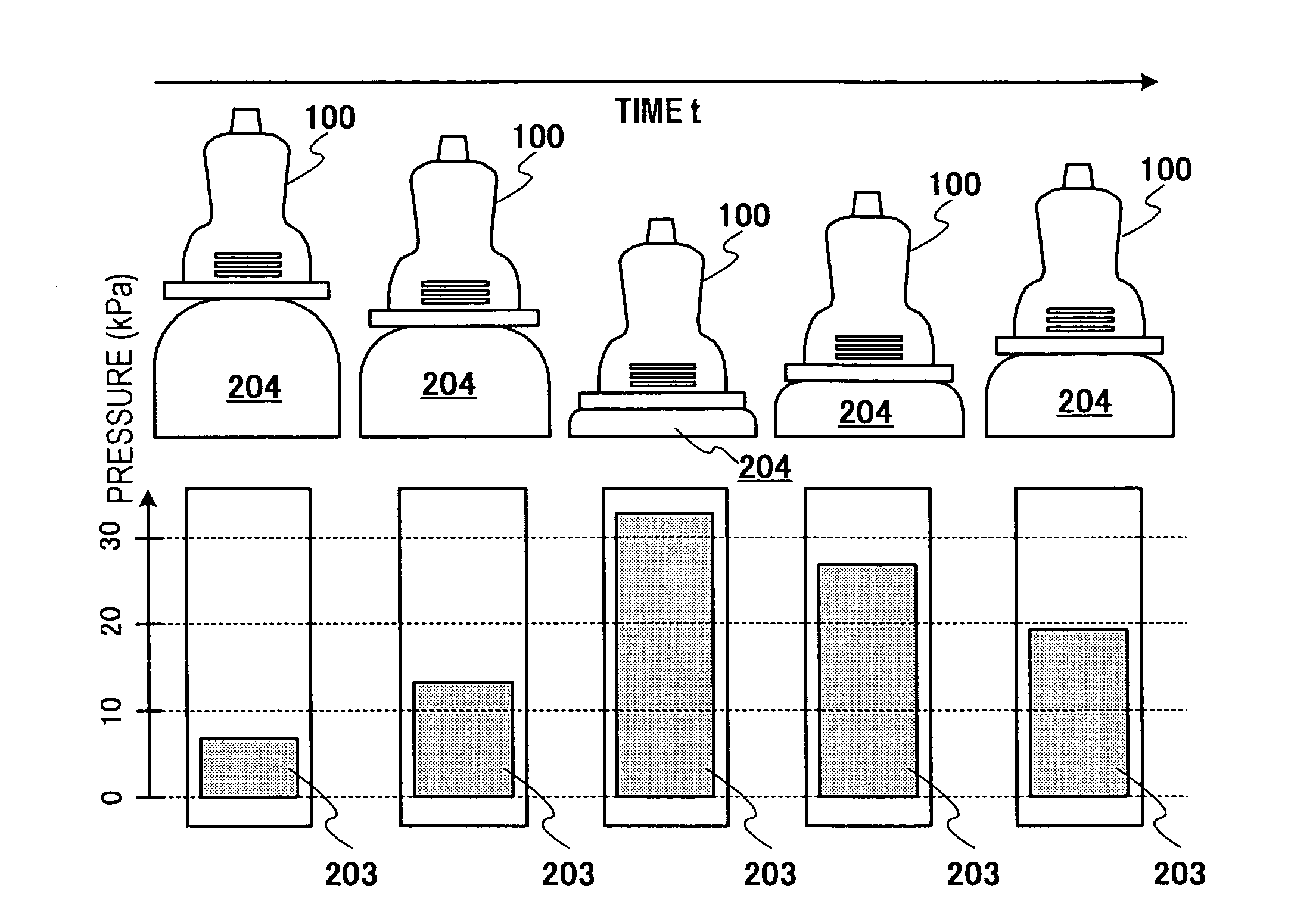
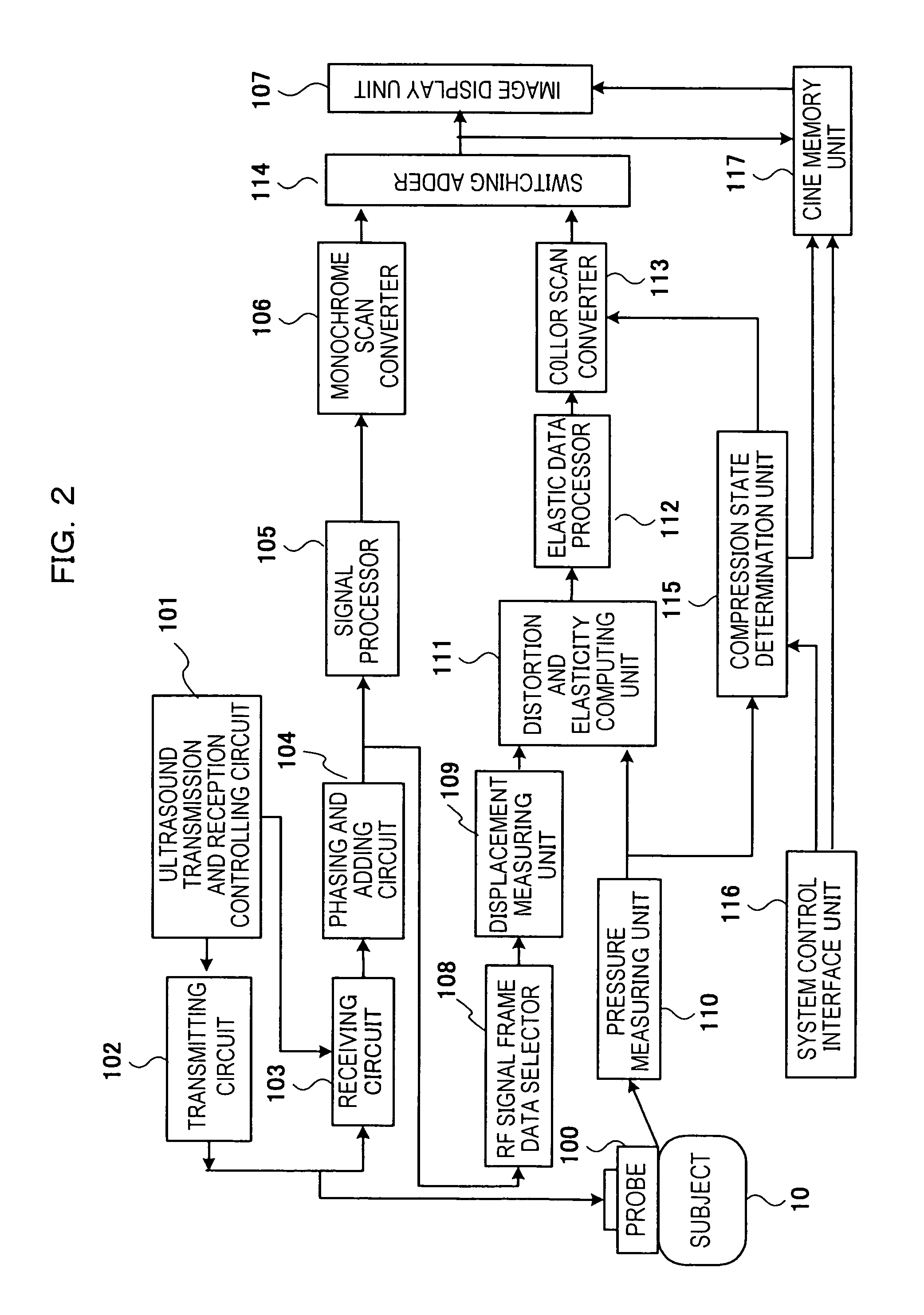
[0079]FIG. 5 is a block diagram illustrating the flow of a process carried out by the compression state evaluating unit 115 according to this embodiment. The compression state evaluating unit 115 includes a memory circuit 1151, a compression state evaluating circuit 1152, and an image configuring circuit 1153. The memory circuit 1151 stores pressure data of the measurement result output from the pressure measuring unit 110 and outputs it to the compression state evaluating circuit 1152. The compression state evaluating circuit 1152 receives the pressure data output from memory circuit 1151, carries out statistical processing, such as overflow processing and average value calculation, on the pressure data so that an optimal image is displayed when displaying the compression state, and outputs numerical value data obtained as a result as compression state evaluation data to the following image configuring circuit 1153. The image configuring circuit 1153 receives the compression state ...
second embodiment
[0101]In the first embodiment, a case in which a pressure data group output from a plurality of pressure sensors in the pressure measuring unit 110 is used to display a statistic characteristic as compression state information is described. However, the present invention is not limited, and sets of pressure data of the pressure sensors may be used to configure sets of compression state image data, and each set of compression state image data may be displayed independently. In other words, when a plurality of pressure sensors is disposed along the long axis direction of the ultrasound transmission and reception surface, as shown in FIG. 6, the positions of the pressure sensors and the corresponding positions in the elastic image and the cross-sectional image may be matched, and each set of compression state image data 10A that is configured independently for each set of the pressure data may be displayed, for example, as shown in FIG. 10. In FIG. 10, the heights of the bar graphs of ...
third embodiment
[0103]FIG. 11 illustrates an example in which the bar graphs in the displayed image in FIG. 9 is replace with a graphic representation 208 simulating the state of compression and deformation of the cross-section region being compressed by the probe shown in the upper section of FIG. 8. By displaying the state of the target tissue being compressed by the probe, the examiner can intuitively recognize the compression state. In FIG. 11, a pressure data value 209 is displayed at the lower area of the schematic diagram of the probe and the target tissue.
PUM
 Abstract
Description
Claims
Application Information
 - R&D
- Intellectual Property
- Life Sciences
- Materials
- Tech Scout
- Unparalleled Data Quality
- Higher Quality Content
- 60% Fewer Hallucinations
Browse by: Latest US Patents, China's latest patents, Technical Efficacy Thesaurus, Application Domain, Technology Topic, Popular Technical Reports.
© 2025 PatSnap. All rights reserved.Legal|Privacy policy|Modern Slavery Act Transparency Statement|Sitemap|About US| Contact US: help@patsnap.com


