
Method and system for human vision model guided medical image quality assessment

a human vision model and image quality technology, applied in the field of human vision model guided image quality assessment, can solve the problems of image quality assessment, difficult to achieve, and difficult to achieve, and achieve the effect of improving the quality of medical image quality assessment, and improving the quality of medical image assessmen

Inactive Publication Date: 2011-12-27
SIEMENS HEALTHCARE GMBH
View PDF8 Cites 18 Cited by
  • Summary
  • Abstract
  • Description
  • Claims
  • Application Information

AI Technical Summary

Problems solved by technology

Image quality assessment is an important and challenging problem in image processing.
However, because of the differences is the users, image formats, and characteristics of image contents in medical images, directly applying existing natural image quality evaluation methods for evaluating the image quality of medical images does not typically provide satisfactory results.
Unlike general users for natural images, physicians typically focus on particular regions of interest (ROI) in a medical image, and the quality of other regions in the image may be irrelevant for diagnostic accuracy.
The formats and characteristics of medical images also make the problem of medical image quality assessment quite different from that of natural images.
However, the original images may not be available or reliable in many situations.
Furthermore, research has shown that traditional quantitative image quality metrics, such as peak signal to noise ratio (PSNR) and mean squared error (MSE), are not directly related to human perception.

Method used


Image

  • Method and system for human vision model guided medical image quality assessment
  • Method and system for human vision model guided medical image quality assessment
  • Method and system for human vision model guided medical image quality assessment

Examples


Embodiment Construction

[0020]The present invention relates to a method for medical image quality assessment. Although embodiments of the present invention are described herein using x-ray images, the present invention can be applied to all types of medical images, such as computed tomography (CT), magnetic resonance (MR), ultrasound, etc. Embodiments of the present invention are described herein to give a visual understanding of the medical image quality assessment method. A digital image is often composed of digital representations of one or more objects (or shapes). The digital representation of an object is often described herein in terms of identifying and manipulating the objects. Such manipulations are virtual manipulations accomplished in the memory or other circuitry / hardware of a computer system. Accordingly, is to be understood that embodiments of the present invention may be performed within a computer system using data stored within the computer system.

[0021]Embodiments of the present inventio...


PUM


Abstract

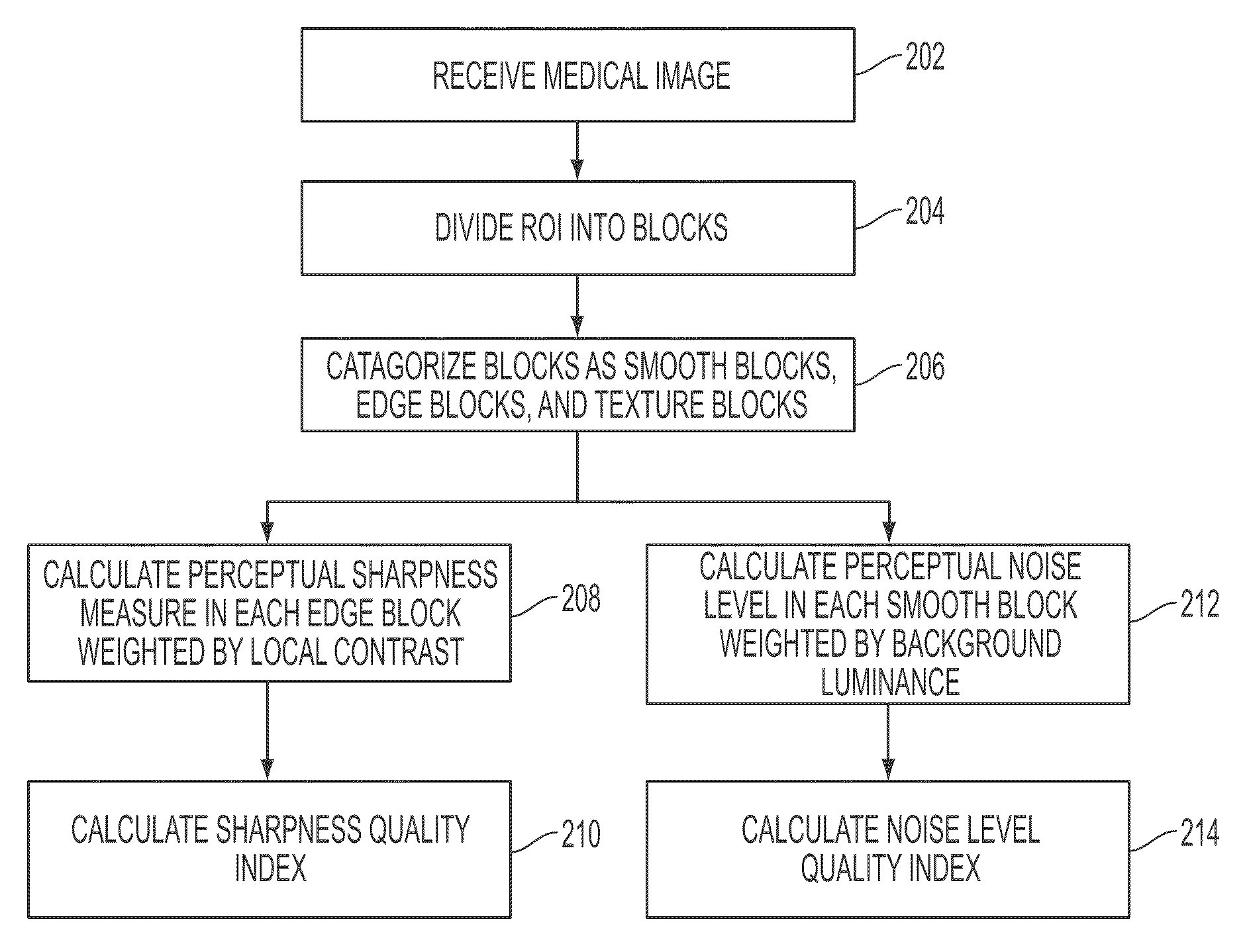
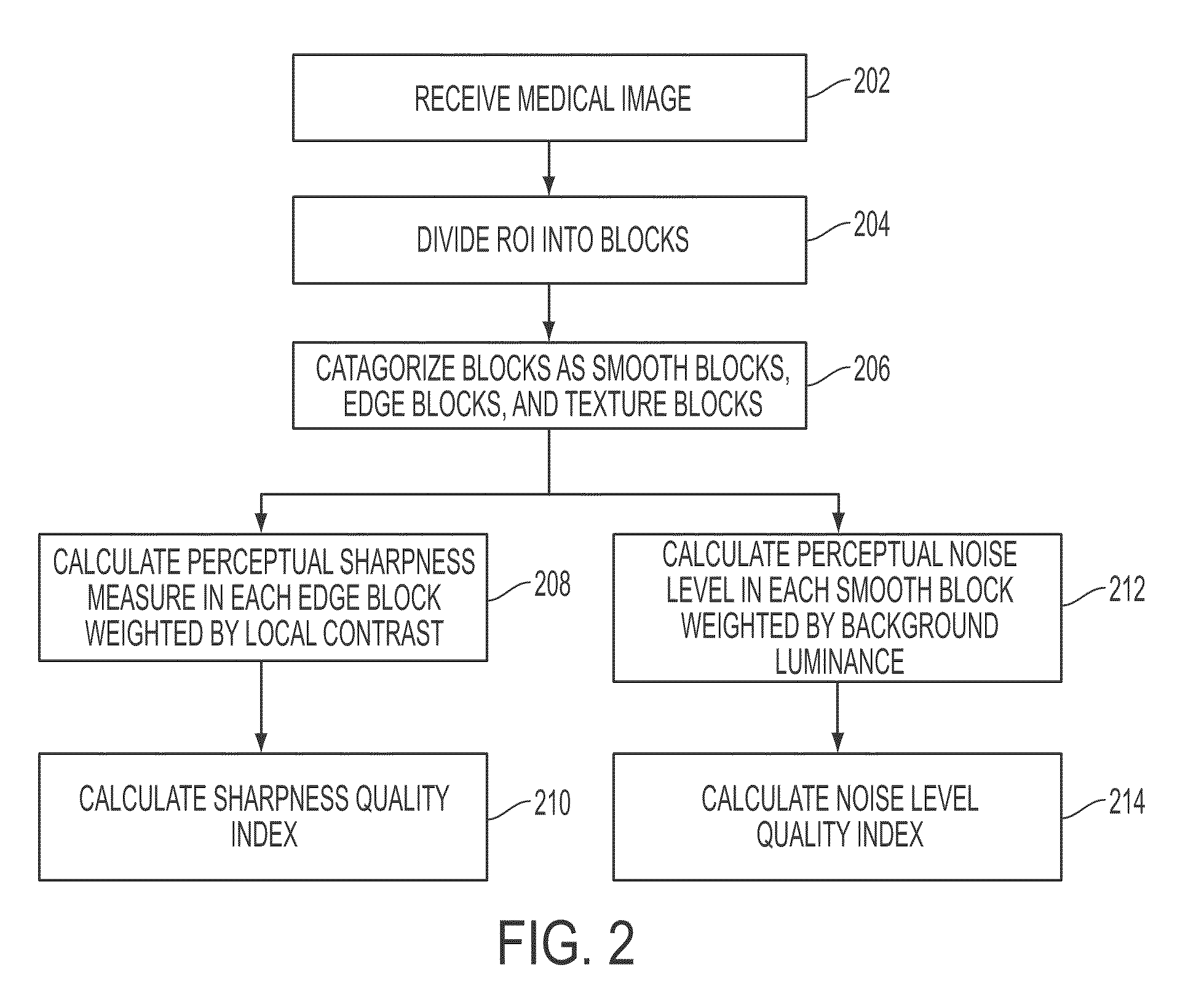
A method and system for image quality assessment is disclosed. The image quality assessment method is a no-reference method for objectively assessing the quality of medical images. This method is guided by the human vision model in order to accurately reflect human perception. A region of interest (ROI) of medical image is divided into non-overlapping blocks of equal size. Each of the blocks is categorized as a smooth block, a texture block, or an edge block. A perceptual sharpness measure, which is weighted by local contrast, is calculated for each of the edge blocks. A perceptual noise level measure, which is weighted by background luminance, is calculated for each of the smooth blocks. A sharpness quality index is determined based on the perceptual sharpness measures of all of the edge blocks, and a noise level quality index is determined based on the perceptual noise level measures of all of the smooth blocks. An overall image quality index can be determined by using task specific machine learning of samples of annotated images. The image quality assessment method can be used in applications, such as video / image compression and storage in healthcare and homeland security, and band-width limited wireless communication.

Description

[0001]This application claims the benefit of U.S. Provisional Application No. 60 / 980,864, filed Oct. 18, 2007, the disclosure of which is herein incorporated by reference.BACKGROUND OF THE INVENTION[0002]The present invention relates to image quality assessment, and more particularly, to human vision model guided image quality assessment in medical images.[0003]Image quality assessment is an important and challenging problem in image processing. With the rapid advancements of digital image processing, the demand of objective image quality assessment is very high. For example, for image compression, an objective image quality metric that measures the quality of a compressed image can be used to find the best compromise between the rate and distortion in the image compression. Image quality evaluation can also be applied to image communication. When transmitting an image progressively, an objective image quality metric can be used to determine if the transmitted image information is g...

Claims


Application Information

Patent Timeline
Patent Type & Authority Patents(United States)
IPC IPC(8): G06K9/00
CPCG06K9/4619G06T7/0012G06K2209/05G06V10/449G06V2201/03
Inventor YAN, MICHELLE XIAO-HONGCHANG, TI-CHIUNLENDL, MARKUSBOEHM, STEFANFANG, TONGDURLAK, PETER
Owner SIEMENS HEALTHCARE GMBH
Learn More