Methods and devices for producing the parameters of the brain tissues and assessing data of the suitability for thrombolysis of a patient

a brain tissue and parameter technology, applied in the field of medical imaging technology, can solve the problems of cell death in minutes, severe ionic disruption and metabolic failure, and zones within flow-compromised territory, so as to save critical time, shorten imaging time, and reduce patient cost

Active Publication Date: 2012-02-21
SHENZHEN INST OF ADVANCED TECH
View PDF2 Cites 5 Cited by
  • Summary
  • Abstract
  • Description
  • Claims
  • Application Information

AI Technical Summary

Benefits of technology

[0012]The object of the present invention is to provide a method and a device for producing the parameters of the brain tissue of a patient quickly by using suitable magnetic resonance imaging, utilizing image processing and computer technology, achieving parameters pertaining to the infarction and the ischemic penumbra.
[0019]There is no need to perform perfusion-weighted imaging (PWI) which is time consuming and invasive. This will shorten the imaging time and save the critical time for decision making, reduce the patient cost and decrease the physical pain of the patient.

Problems solved by technology

Within the centre or core of the ischemic territory, blood flow deficits, low ATP (adenosine triphosphate) levels and energy stores, ionic disruption and metabolic failure are severe, and cell death progresses in minutes.
However, the peripheral zones within the flow-compromised territory—the ischemic penumbra—suffer milder insults due to residual perfusion from collateral blood vessels.
Within the core territory, salvage of rapidly dying brain cells might not be feasible without early reperfusion.
However, due to its limited availability and long post-processing time, PET cannot be used clinically to guide acute stroke therapy.
However, it is widely recognized that the mismatch model is at best a crude approximation of the ischemic penumbra because 1) the perfusion techniques and data evaluation are not truly quantitative and vary among centers; 2) magnetic resonance PWI is invasive and time consuming, and has not been approved by health authorities; 3) the thresholds used for defining perfusion abnormalities have not been validated against PET thresholds; and 4) abnormal diffusion regions early after stroke onset can be reversed by timely intervention in both animals and humans.
Though appropriate thrombolysis can greatly improve prognosis of patients and save lives, inappropriate thrombolysis could cause death and symptomatic brain hemorrhage.
Even with the only 2 reported successful intravenous thrombolysis stroke trials by NINDS (National Institute of Neurological Disorders and Stroke) within 3 to 6 hours after symptom onset, there were 10-fold increased risk of symptomatic brain hemorrhage guided by the mismatch model.

Method used


Image

  • Methods and devices for producing the parameters of the brain tissues and assessing data of the suitability for thrombolysis of a patient
  • Methods and devices for producing the parameters of the brain tissues and assessing data of the suitability for thrombolysis of a patient
  • Methods and devices for producing the parameters of the brain tissues and assessing data of the suitability for thrombolysis of a patient

Examples


example 1

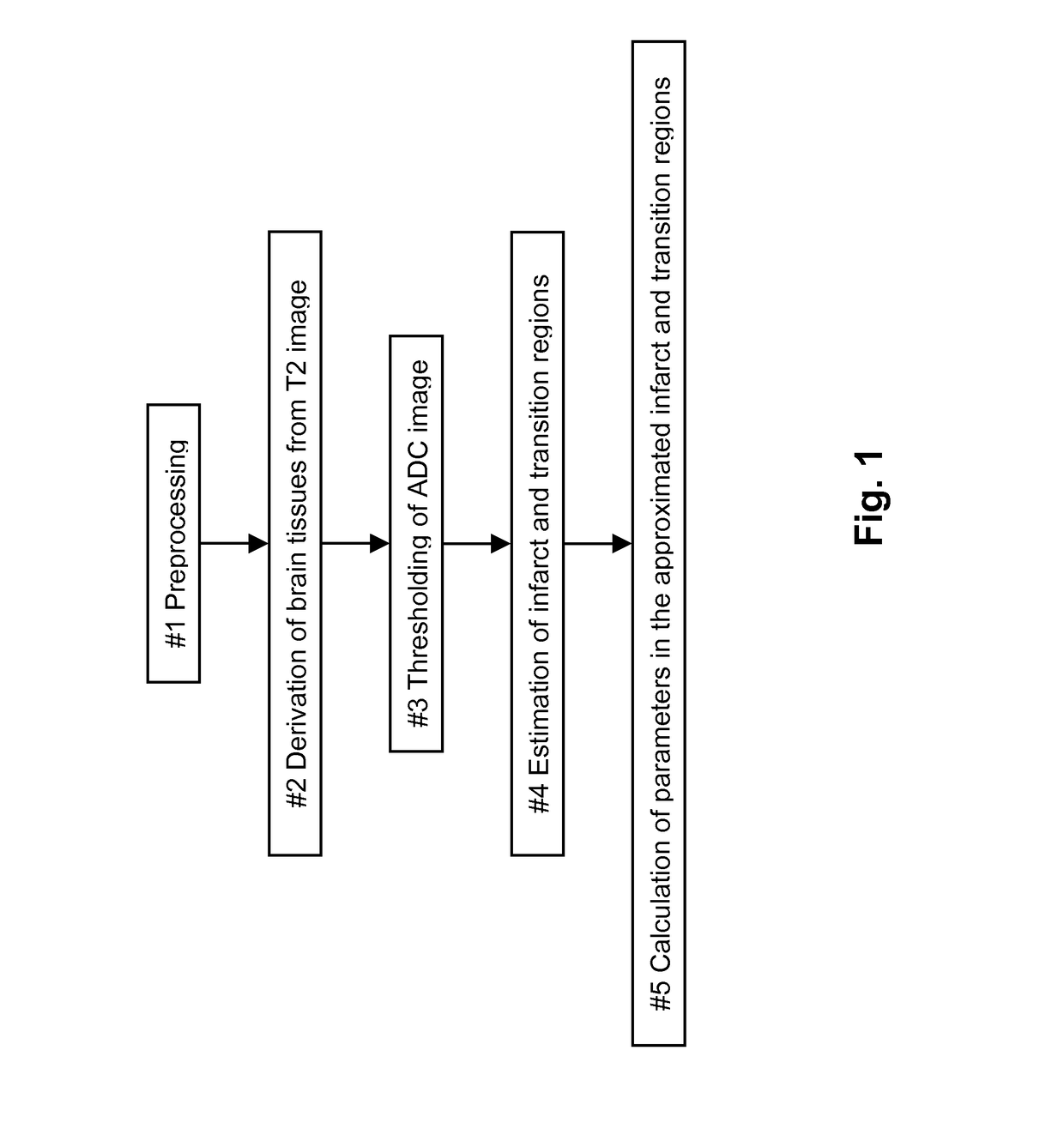
The Method for Producing the Parameters of the Brain Tissues of a Patient

[0035]The method of the present invention utilizes the Diffusion-Weighted Imaging (DWI) of a ischemic stroke patient, including two volumetric data form diffusion weighted imaging: (1) T2-weighted image (i.e., the DWI with the gradient factor b being 0) denoted as T2(x, y, z), and (2) the ADC image denoted as ADC(x, y, z)), both of which have the same voxel sizes, same coordinate systems, and the same coordinates correspond to the same physical positions, obtained when the patient is admitted to have the acute DWI scan.

[0036]Both T2(x, y, z) and ADC(x, y, z) are in 16-bits DICOM format. It is assumed that the voxel sizes in the X, Y, and Z directions are denoted as voxX, voxY, and voxZ, respectively with mm / voxel as their units. Here (x, y, z) are integer coordinates of voxels satisfying 0≦x≦xSize-1, 0≦y≦ySize-1, 0≦z≦zSize-1. The z coordinate is constant on axial, y on coronal, and x on sagittal slices.

[0037]A ...

example 2

The Medical Device Producing the Parameters of the Brain Tissues of a Patient, the Device Comprising

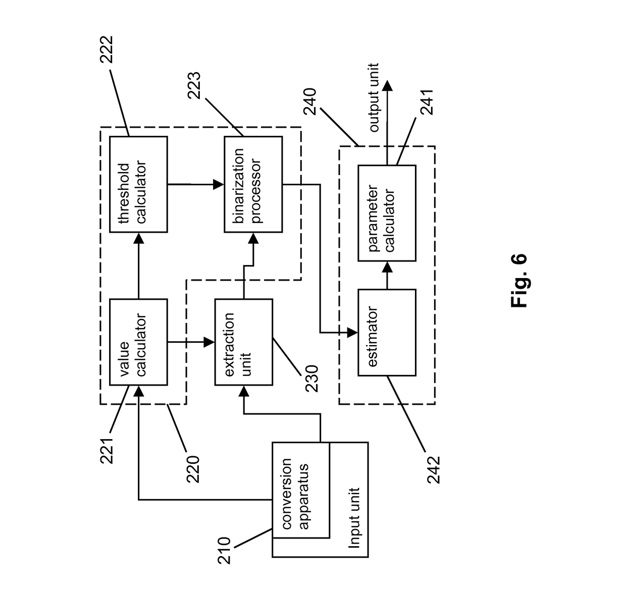
[0093]Based on the method described above, a medical device comprises at least an input unit 210 and a processing module. The input unit provides interface for inputting the DICOM data of the patients T2(x, y, z) and ADC(x, y, z), and the processing module to perform the processing described in steps 1 to 5.

[0094]The input unit 210 has a conversion apparatus converting the image from 16-bit data to 8-bit or 4-bit data, i.e. an apparatus implementing the step #1.

[0095]The processing module comprises an extraction unit 230 extracting a brain tissues image from the T2-weighted image, an image processing unit 220 achieving a core ADC threshold and a transition ADC threshold that could differentiate a core region and a transition region respectively and binarizing the ADC image through the transition ADC threshold according to the brain tissues image to achieve a binarized ADC image, and a...

example 3

The Method for Producing and Assessing Data of the Suitability for Thrombolysis of a Patient

[0101]The method of the present invention comprises a group of steps in which the first step to the five step are the same as example 1 and the sixth step to the seven step are as follows:

[0102]Step #6: Assessing if There Exist Ischemic Tissues to be Rescued

[0103]To assess if there exist ischemic tissues to be rescued is to see if the transition regions contain ischemic tissues to be rescued. This is achieved through training samples. A patient's data is used as one of the training samples if it meets one of the following criteria: 1) the patient shows substantial improvement after thrombolysis, which implies that this patient has ischemic tissues to be rescued and these tissues have been rescued through thrombolysis; 2) the patient symptoms are deteriorated after thrombolysis, which implies that this patient should not be handled with thrombolysis; and 3) those patients that show no mismatch...


PUM


Abstract

This invention provides a method and a device for producing the parameters of the brain tissue of a patient quickly by using suitable magnetic resonance imaging, utilizing image processing and computer technology, achieving parameters pertaining to the infarction and the ischemic penumbra. This invention also provides a method and a device for producing and assessing data of the suitability for thrombolysis of a patient quickly and properly by using suitable magnetic resonance imaging, utilizing image processing and computer technology, achieving parameters pertaining to the infarction and the ischemic penumbra and predicting the suitability for thrombolysis.

Description

FIELD OF THE INVENTION[0001]The present invention relates to a medical imaging technology, in particular, relates to a method and a device for producing the parameters of the brain tissues of a patient utilizing magnetic resonance imaging (MRI).[0002]The present invention also relates to a method and a device for producing and assessing data of the suitability for thrombolysis utilizing magnetic resonance imaging (MRI).BACKGROUND OF THE INVENTION[0003]According to the Chinese Ministry of Health, stroke is the second cause of mortality and the first cause of disability in China. There are about 600,000 to 700,000 stroke patients in China, 130,000 to 150,000 new stroke patients and about 100,000 patients died of stroke each year. Among those who survived from the initial symptom onset, about 3 quarters of the patients are disabled to some extents, with about 40% being seriously disabled. Each year China spends more than 30 billion RMB for stroke health care.[0004]According to the Amer...

Claims


Application Information

Patent Timeline
Patent Type & Authority Patents(United States)
IPC IPC(8): G06K9/00
CPCA61B5/055G01R33/56341G06T7/0012G01R33/5608G06T2207/10088G06T2207/20148G06T2207/30016A61B5/4076G06T7/136
Inventor HU, QINGMAO
Owner SHENZHEN INST OF ADVANCED TECH
Why Eureka
Try Eureka