Oncolytic virus

a technology of oncolytic virus and oncolytic peptide, which is applied in the field of oncolytic virus, can solve the problems of limited use of oncolytic virus common human pathogens, and the possibility of tumor regrowth

Inactive Publication Date: 2012-04-03
WELLSTAT BIOLOGICS CORP
View PDF42 Cites 4 Cited by
  • Summary
  • Abstract
  • Description
  • Claims
  • Application Information

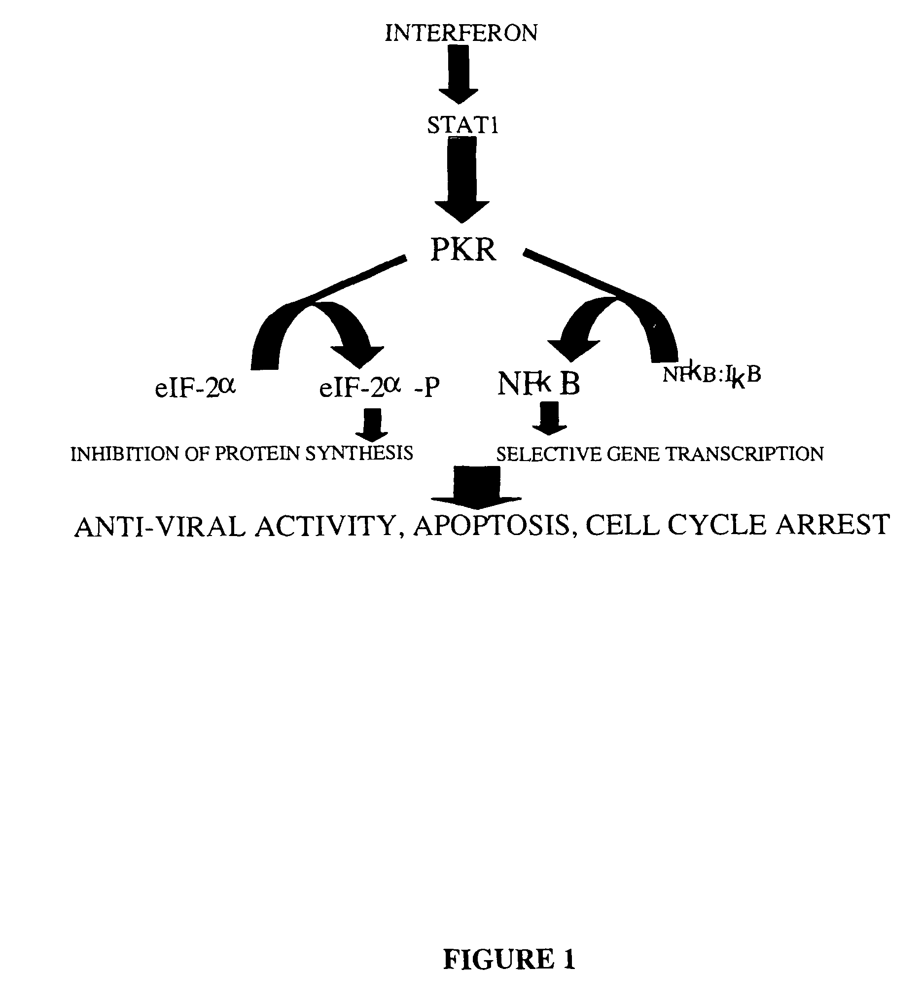
AI Technical Summary

Benefits of technology

[0025]There are several advantages for the use of a virus as described herein as a therapeutic virus over other viruses:
[0026]Rhabdoviruses are not common human pathogens. For example, VSV is found mostly in insects, rodents and domestic farm animals, and therefore a large proportion of individuals will not have been infected or immunized to VSV infection. On the other hand, Adenovirus or Reovirus are human pathogens and most of the general population have been infected and acquired an immune response to both of these viruses. A preexisting immune response to a viral strain similar to the one used as a therapeutic agent in the treatment of a cancer may attenuate the effectiveness of the virus as therapeutic agent;
[0029]Cellular infection by VSV is highly responsive to additional chemical agents such as interferon, a feature which enhances its therapeutic value.
[0031]VSV is a RNA virus and spends its entire lifecycle in the cytoplasm. Therefore it involves less danger of unwanted integration into the genome of a patient.Collectively, these VSV attributes provide significant advantages over the use of the other viruses known to exhibit oncolytic activity.

Problems solved by technology

However in many instances, for example with the use of Clostridium, the tumours are only partially destroyed, and tumour regrowth may still occur.
Furthermore, the use of common human pathogens as an oncolytic virus is limited as it is likely that the general population has been infected and acquired an immune response to such viruses.

Method used


Image

  • Oncolytic virus
  • Oncolytic virus
  • Oncolytic virus

Examples


example 1

PKR Negative Cells are Susceptible to VSV Infection

In Vivo Experiments

[0111]Initial studies were directed to identifying viruses that are capable of infecting PKR− / − animals and cells. Using homologous recombination strategies, PKR null mouse strains were generated (35, which is incorporated by reference) and tested for their ability to fight virus infections. Since these mice are PKR− / −, they should be susceptible to virus infection. Several species of virus were administered to PKR null animals over a range of concentrations.

Infection of PKR Null Mice:

[0112]A PKR null mouse line was generated using conventional knockout technology (Abraham, N., et al., J Biol Chem, 1999. 274:5953-5962). Groups of five female mice, 3 months of age or greater, were infected intranasally with varying amounts of vesicular stomatitis virus (Indiana strain). Age matched wild type animals were infected in parallel and both sets of animals were monitored on a daily basis for signs of infection. These incl...

example 2

Selective Killing of Tumour Cells with VSV

In Vitro Experiments

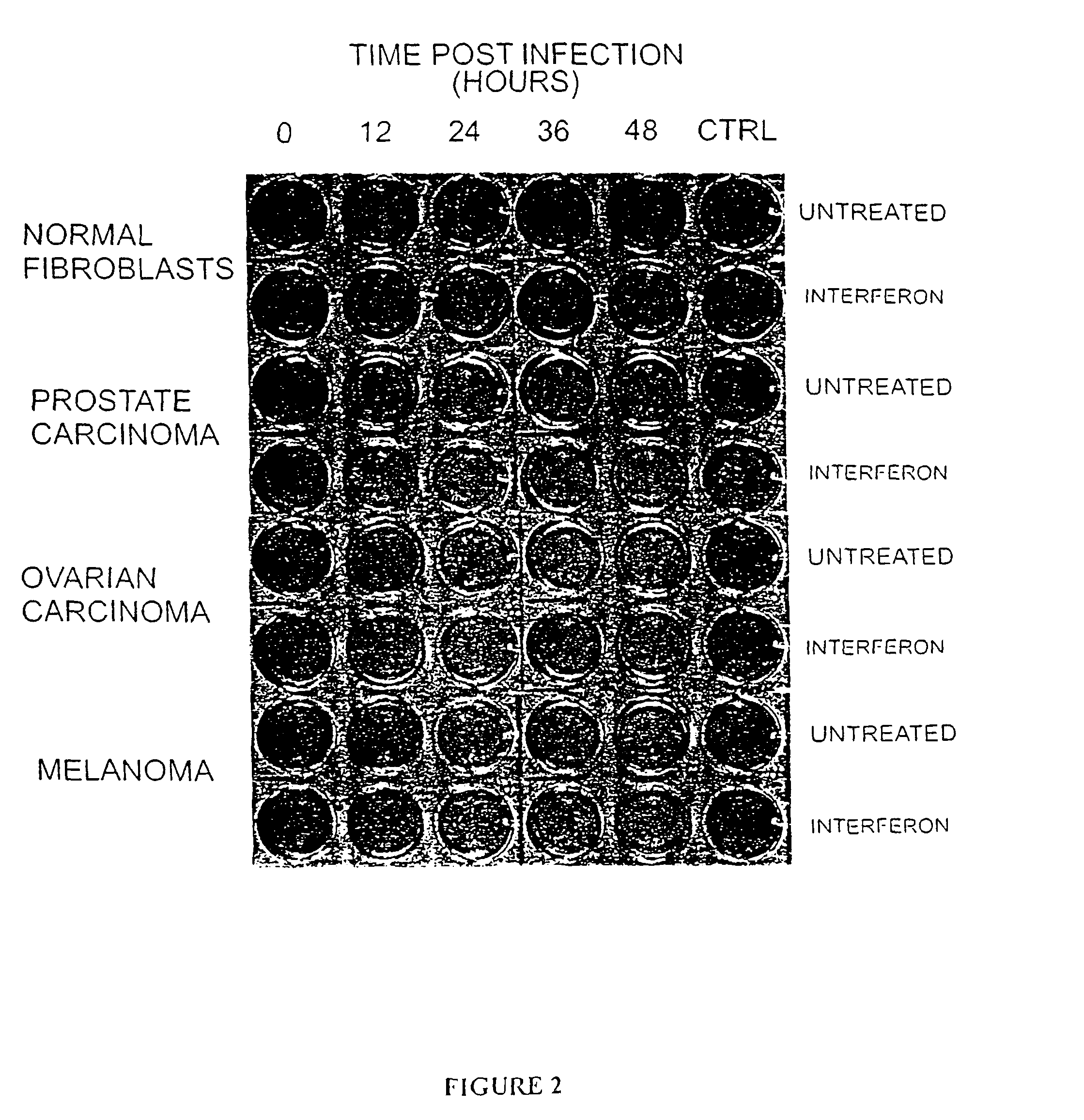
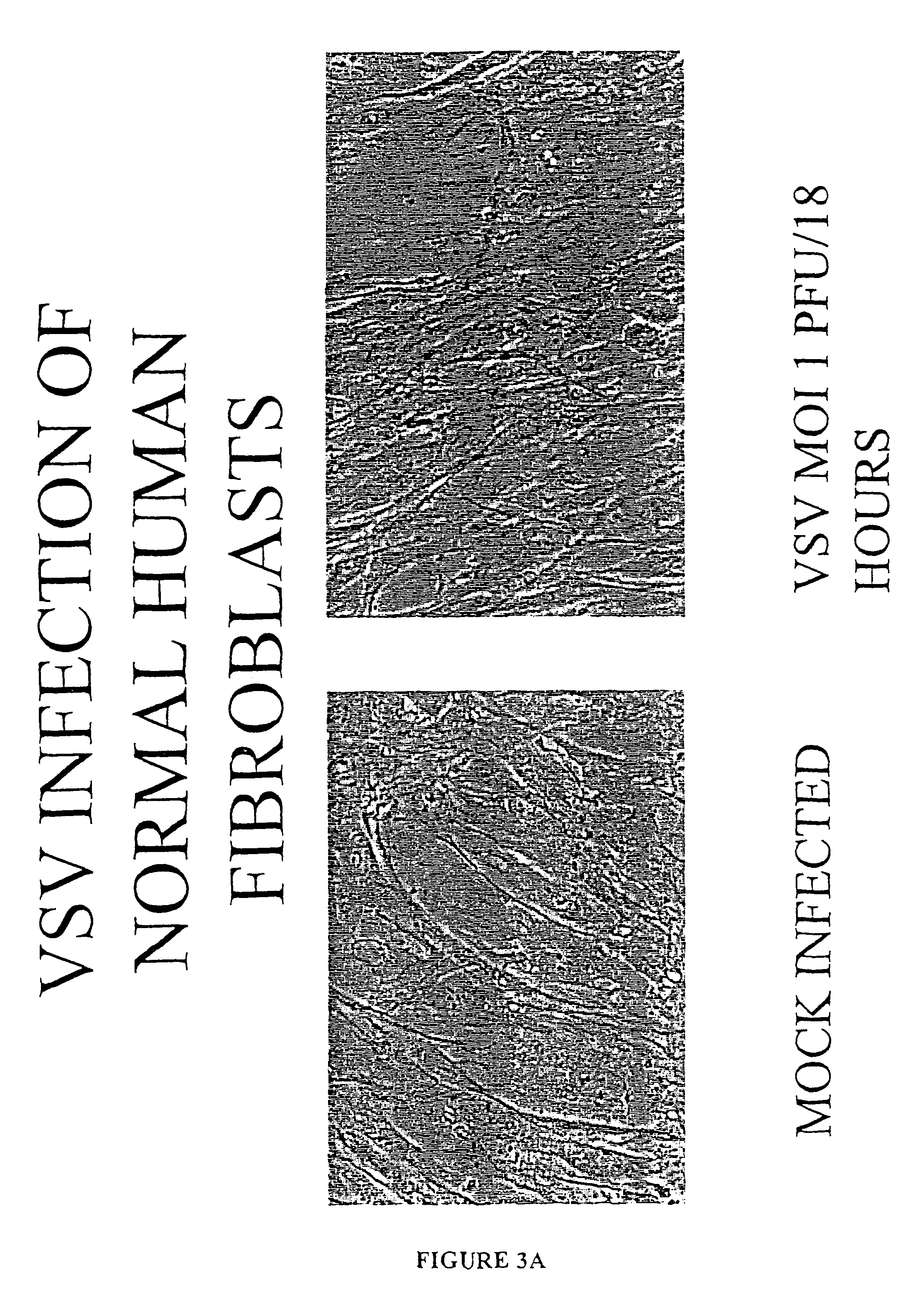
[0115]Several tumour cell lines were chosen at random from the Ottawa Regional Cancer Center and tested for their susceptibility to VSV infection. Primary fibroblast cultures from healthy adult volunteers or primary bone marrow samples from healthy donors were used as control cells.

Infection of Tumour Cells with VSV:

[0116]As a first test of the oncolytic properties of VSV, virus production and cytopathic effect following an overnight incubation with VSV was assessed. Monolayers of cells were incubated with the Indiana strain of VSV at a multiplicity of infection (moi) of 0.1 plaque forming units (pfu). After allowing virus to adsorb for minutes at 37 C, the cultures were rinsed thoroughly with phosphate buffered saline (PBS) and then cultured an additional 18 hours at 37 C. At this time, the cultures were examined microscopically for cytopathic effect (cpe) and photographed. The 18 hour supernatant was removed and virus t...

example 3

Killing of Tumour Cells in Mixed Cultures

[0125]Normal human fibroblasts and 293T tumour cells were co-cultured in a 50:50 mixture. Since 293T cells express the large T antigen which is not found in normal cells, the two cell types can be distinguished by immunofluoresence.

[0126]In this experiment cultures were infected at an moi of 0.1 pfu / cell and the infection allowed to proceed in the presence or absence of interferon. At 0, 18 and 24 hours (FIG. 4) the cultures were fixed and stained with antibodies to large T antigen (red nuclei) to detect the 293T cells and with DAPI (blue nuclei) which stains all cell types (FIG. 4). Initially both cell types displayed a spindle-like morphology with large oval nuclei. After 18 hours the number of 293T cells (red nuclei) were reduced and many of the remaining 293T cells displayed altered nuclear morphology. By 24 hours post-infection very few 293T cells were detected and those few that remained displayed severely condensed or fragmented nuclei...


PUM

PropertyMeasurementUnit
timeaaaaaaaaaa
volumeaaaaaaaaaa
timeaaaaaaaaaa

Abstract

The present invention is directed to a method of reducing the viability of a tumor cell involving administering a virus that is not a common human pathogen to the tumor cell. Preferably, the virus exhibits differential susceptibility, in that normal cells are not affected by the virus. This differential susceptibility is more pronounced in the presence of interferon. The tumor cell is characterized by having low levels, or no, PKR activity, or as being PKR− / −, STAT1− / − or both PKR− / − and STAT1− / −. The virus is selected from the group consisting of Rhabdovirus and picornavirus, and preferably is vesicular stomatitis virus (VSV) or a derivative thereof.

Description

[0001]This application claims the benefit of U.S. Provisional Application No. 60 / 287,590, having an effective filing date of Sep. 17, 1999 now abandoned, as obtained by the petition dated Sep. 8, 2000 under 37 CFR 1.53(c)(2) from U.S. application Ser. No. 09 / 397,873, filed Sep. 17, 1999, the contents of which are incorporated herein by reference.[0002]The present invention relates to a novel cancer therapeutic. More specifically, this invention relates to viruses that selectively infect and inhibit tumour cell growth.BACKGROUND OF THE INVENTION[0003]The use of oncolytic bacteria, or compositions of oncolytic bacterias, for combatting neoplasms in humans and animals is known. For example EP 564 121, GB 1,587,244 and U.S. Pat. No. 3,192,116 disclose the use of non-pathogenic bacteria that result in the liquification and lysis of tumours in vertebrates. However in many instances, for example with the use of Clostridium, the tumours are only partially destroyed, and tumour regrowth may ...

Claims


Application Information

Patent Timeline
Patent Type & Authority Patents(United States)
IPC IPC(8): A61K35/76A61K35/766A61K48/00C07K14/145C12N7/02C12N15/86
CPCA61K35/76A61K35/766C07K14/005C12N7/00C12N15/86A61K35/13A61K48/00C12N2760/20222C12N2760/20232C12N2760/20251C12N2810/6081A61K38/21A61K2300/00
Inventor BELL, JOHN C.SONENBERG, NAHUMSTOJDL, DAVID F.BROWN, EARL G.ATKINS, HAROLD L.MARIUS, RICARDO M.LICHTY, BRIAN D.KNOWLES, SHANE B.
Owner WELLSTAT BIOLOGICS CORP
Why Eureka
Try Eureka