Radiotherapy and imaging methods and apparatus

a radiotherapy and patient technology, applied in the field of radiotherapy, can solve the problems of significant artifacts in the final image, double quantum noise in the scattering, and interference of therapeutic radiation,

Active Publication Date: 2012-07-10
ELEKTA AB
View PDF10 Cites 31 Cited by
  • Summary
  • Abstract
  • Description
  • Claims
  • Application Information

AI Technical Summary

Benefits of technology

[0007]In contrast, the present invention sums the data acquired in odd and even frames, as this leads to a clean image without artifacts (but with twice the scatter). The summed data can then be processed in order to estimate and remove the scatter from the therapeutic source of radiation.

Problems solved by technology

However, since one aims at minimal dose delivery times, interruption of the therapeutic radiation is not preferred.
The difficulty in this approach is that quantum noise in the scatter is doubled (it does not cancel) and that, due to the non-linear detector “ghosting” signal, significant artifacts occur in the final image.

Method used


Image

  • Radiotherapy and imaging methods and apparatus
  • Radiotherapy and imaging methods and apparatus
  • Radiotherapy and imaging methods and apparatus

Examples


Embodiment Construction

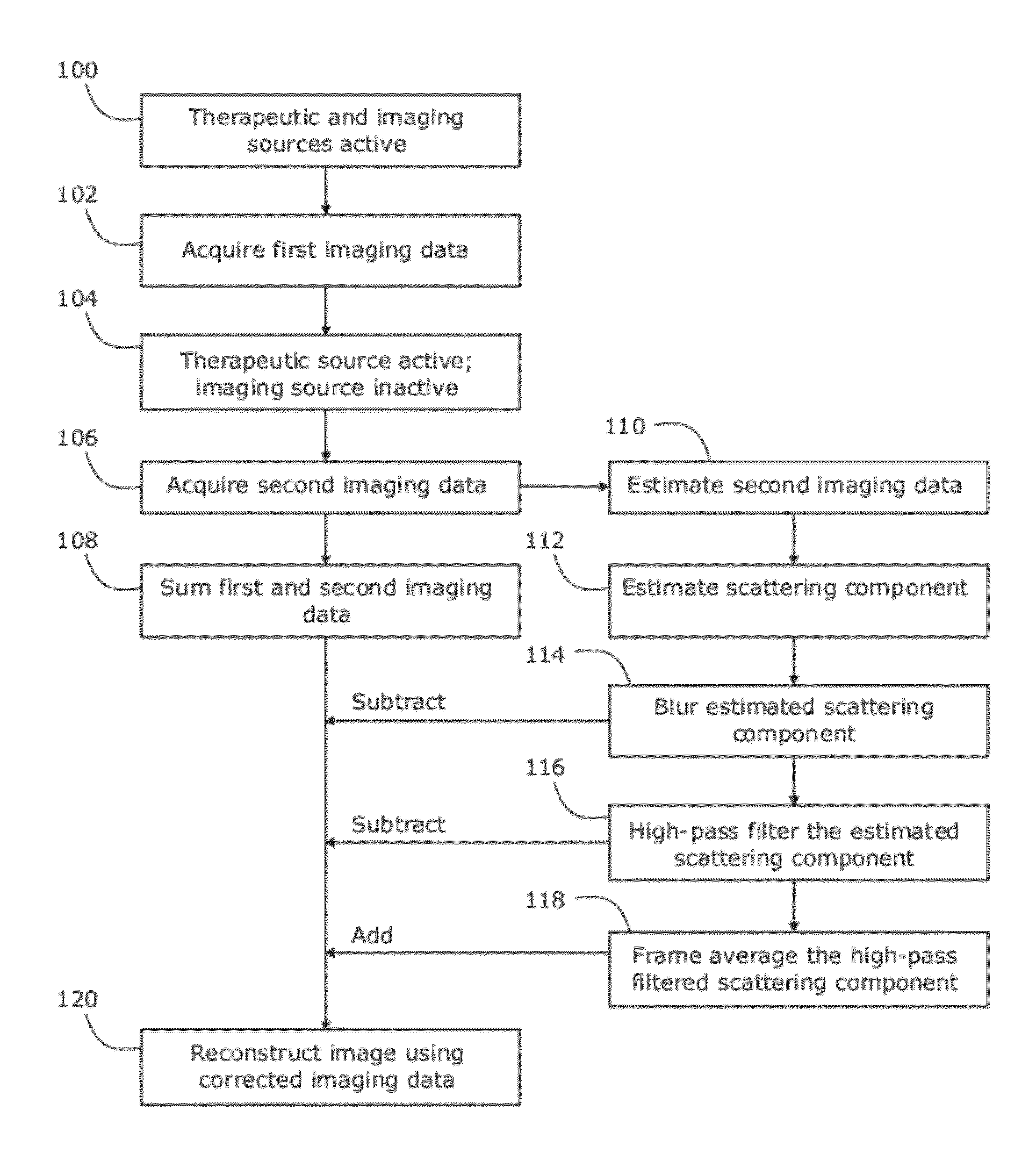
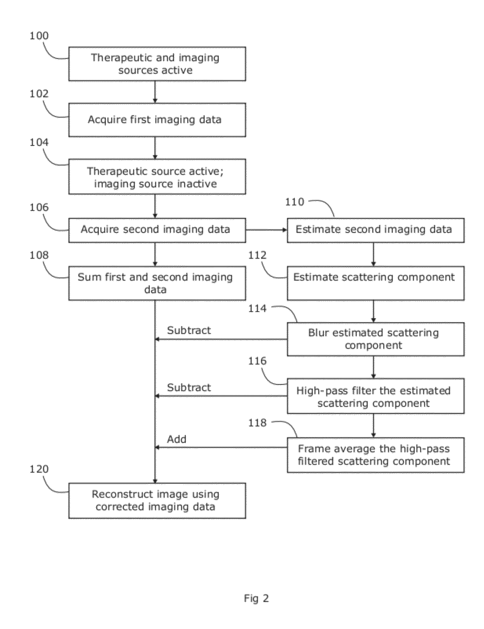
[0014]The present inventors have developed a method and apparatus to correct for MV scatter reaching the kV detector during simultaneous acquisition of CBCT scans with rotational radiotherapy.

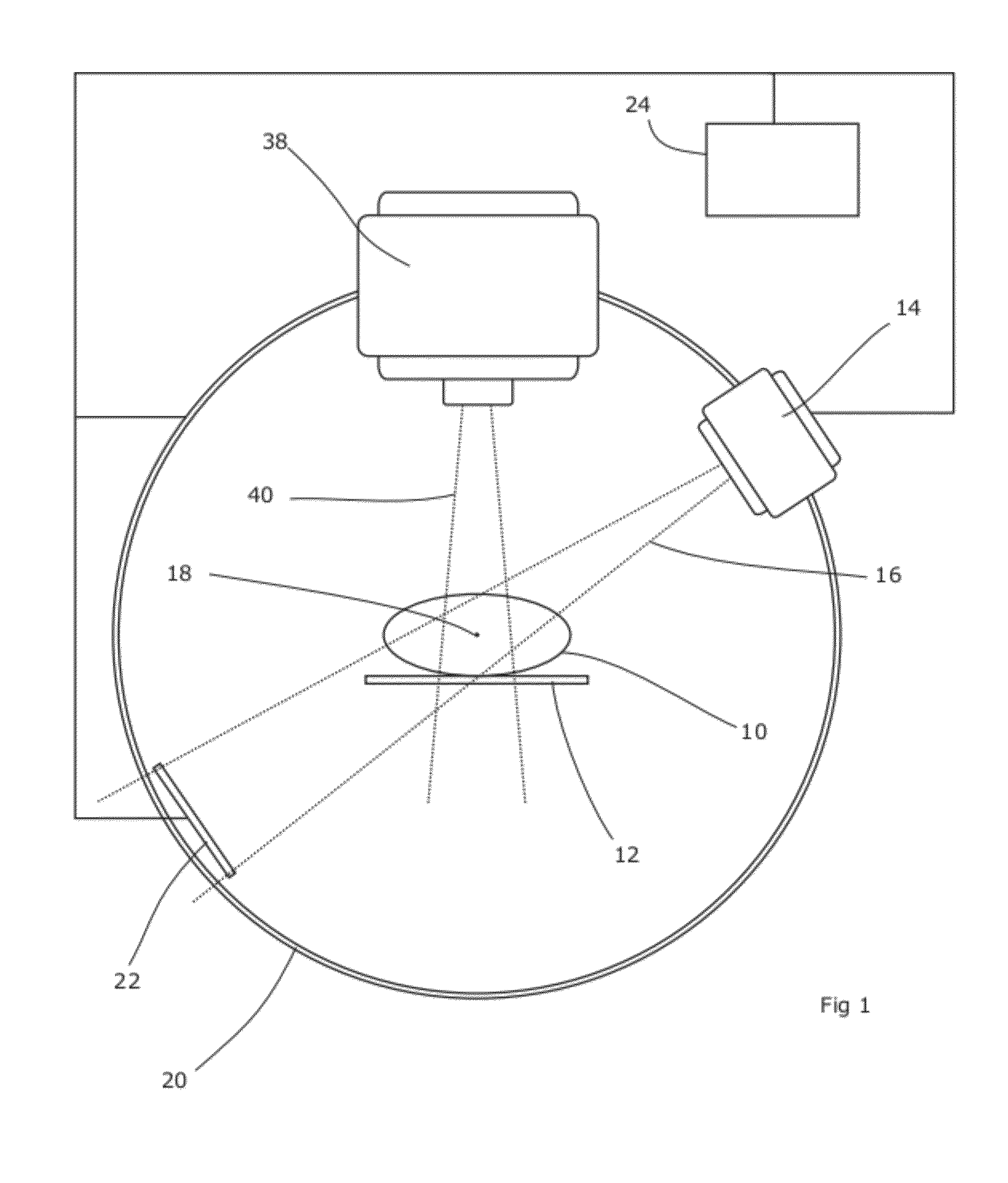
[0015]FIG. 1 is a schematic diagram showing an apparatus according to embodiments of the present invention.

[0016]A patient 10 is supported on a couch 12 which may be of any suitable design. Couches typically allow the elevation, latitudinal and longitudinal position of the patient to be adjusted, and this may be provided for as desired. The couch 12 may also allow rotation in up to three rotational degrees of freedom (pitch, yaw and roll).

[0017]An x-ray source 14 is arranged to project a wide beam 16 of radiation generally directed towards the isocentre 18 of the system. The source 14 is rotatable around the isocentre 18 on a rotational support 20. The support can, for example, be in the form of a ring or annulus around the patient 10 and couch 12 in which the source is mounted, or it can be a ...


PUM


Abstract

The present invention provides a method and an apparatus for reconstructing images from data acquired during radiotherapy. The approach is based on summing the imaging data acquired while both therapeutic and imaging source are active, with that acquired when only the therapeutic source is active. Further correction of the summed data can lead to reconstructed images of excellent quality.

Description

FIELD OF THE INVENTION[0001]The present invention relates to methods and apparatus for imaging a patient undergoing radiotherapy.BACKGROUND ART[0002]Radiotherapy techniques have improved dramatically in recent years. One particular improvement is the integration of radiotherapy with image acquisition devices such as cone-beam computed tomography (CBCT) scanners. In these systems, a moveable (e.g. rotatable) gantry supports a source of therapeutic radiation (i.e. in the megavolt range) as well as a source of imaging radiation (i.e. in the kilovolt range). A detector is positioned on the gantry substantially opposite the source of imaging radiation, and acquires imaging data as the gantry rotates around the patient. Such an imaging device allows efficient data collection for off-line decision protocols, validation of the patient geometry during actual therapy and detection of intra-fraction motion in two dimensions as well as cone-beam computed tomography (CBCT) to verify the patient ...

Claims


Application Information

Patent Timeline
Patent Type & Authority Patents(United States)
IPC IPC(8): A61B6/00A61N5/10
CPCA61N5/1049A61B6/032A61B6/4085A61B6/54A61N2005/1061
Inventor VAN HERK, MARCELSONKE, JAN-JAKOB
Owner ELEKTA AB
Why Eureka
Try Eureka