Method for registering and merging medical image data

a technology of image data and registration, applied in the field of image data registration and superimposition, can solve the problems of increasing the difficulty of guiding the instrument accurately, the difficulty of accurately guiding and the complexity of the guidance of the catheter through the arterial system

Active Publication Date: 2012-08-14
SIEMENS HEALTHCARE GMBH
View PDF14 Cites 10 Cited by
  • Summary
  • Abstract
  • Description
  • Claims
  • Application Information

AI Technical Summary

Benefits of technology

[0004]Guiding the instrument accurately is a challenging task. It is indeed in neuroradiological interventions that the guidance of the catheter though the arterial system can be complicated considerably by the large number of ramifications and convoluted parts of arteries. To assist the physician, it is known for the intervention to be assisted by an imaging system, in particular by an imaging method that uses a mono- or biplanar X-ray system. In such a method, two-dimensional projection images of the region that is being investigated are continually supplied and displayed to the physician during the intervention via the imaging system. The aforementioned images allow the current position of the instrument to be detected. However, orientation within the patient's anatomy becomes more and more difficult because the image detection is two-dimensional.
[0007]Until now the incidence of this problem was reduced by registration of the 2D / 3D image data sets. In this registration, the 3D image data set is adapted to the respective current 2D data set using geometrical operations, in particular translations and rotations. This is achieved by using the information contained in both data sets. One option for registration consists in identifying characteristic structures that are visible in both data sets. The two data sets are then aligned by using these structures. The structures can be anatomical landmarks, for example blood vessels or bones, or artificial markers that are affixed to the patient during the 3D and 2D imaging. A further option for registration is the use of image-based methods that attempt to maximize the similarity between artificially calculated projections from the 3D image data set and the 2D image.
[0012]It is therefore an object of the invention to provide a method for the registration and superimposition of image data when taking serial radio graphs in medical imaging that allows accurate superimposition without time-consuming user interaction.
[0016]The present method allows the registration and superimposition of image data sets to be achieved reliably and in real time, independent of the image quality, when taking serial radiographs in medical imaging. The method is particularly suitable for 2D / 3D image registration and superimposition, particularly in the area of road mapping during a medical intervention. Registration of so-called 4D data sets, that is, of data sets that also contain time data in addition to three-dimensional position data, with other data sets is likewise possible using the present method. Since the method dispenses with artificial or anatomical markers it does not require time-consuming user interaction during the patient's treatment. The only additional step involved optionally consists in installing the position sensor for the location system and likewise optionally in calibrating said location system on one occasion. The calibration is very simple, however, and can easily be combined with the calibration for the 3D data acquisition. The proposed method for registration of the image data using the location system does not require any time-consuming computing operations and can thus be easily carried out in real time.

Problems solved by technology

Guiding the instrument accurately is a challenging task.
It is indeed in neuroradiological interventions that the guidance of the catheter though the arterial system can be complicated considerably by the large number of ramifications and convoluted parts of arteries.
However, orientation within the patient's anatomy becomes more and more difficult because the image detection is two-dimensional.
If the patient moves between the construction of the 3D image data set and the subsequent drawing of a 2D image, the calibration is no longer valid.
In the case of neuroradiological interventions, this can, for example, result in the position of the catheter being shown incorrectly in the 3D images.
As a result of the fact that the dose of radiation that can be applied is limited, and that the amount of contrast agent that can be applied is limited, such recalibrations cannot be carried out frequently, however.

Method used


Image

  • Method for registering and merging medical image data
  • Method for registering and merging medical image data
  • Method for registering and merging medical image data

Examples


Embodiment Construction

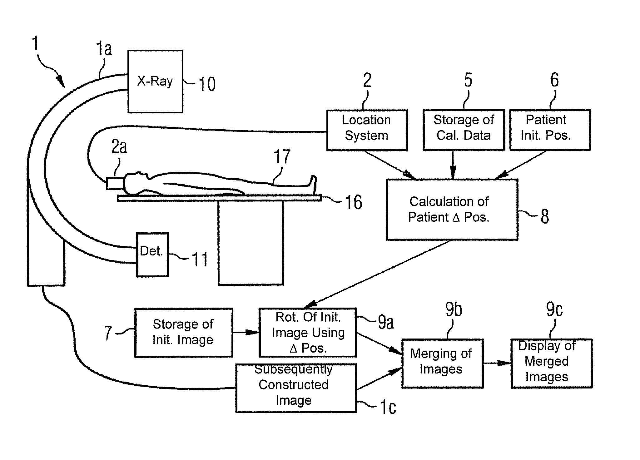
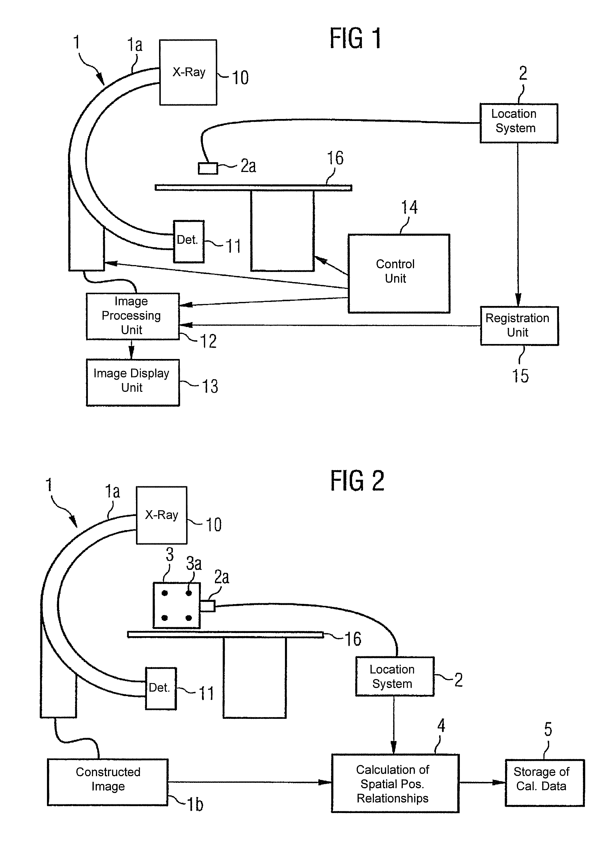
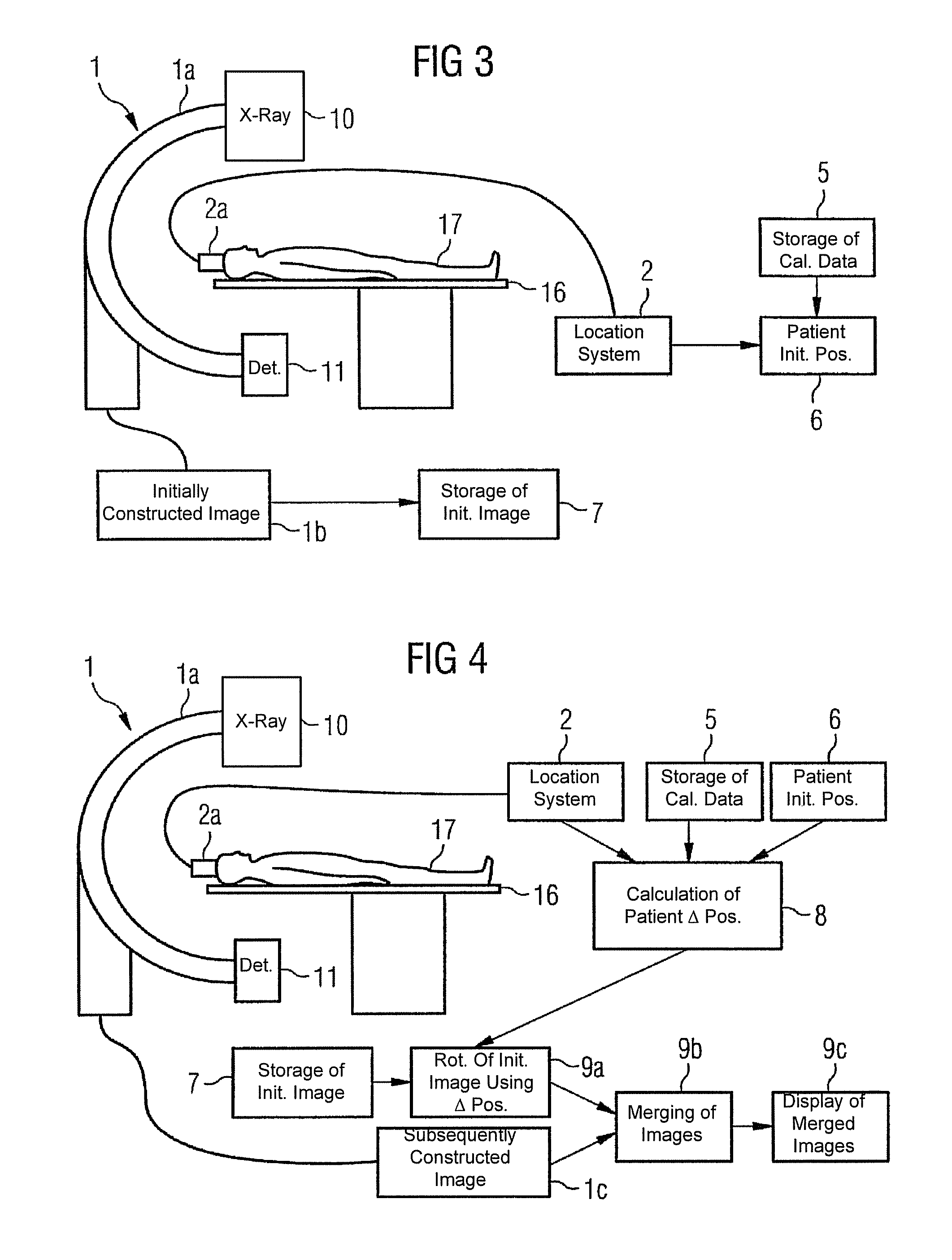
[0024]The present method is hereafter described by means of an x-ray angiography unit for applications in neuroradiology. The method can of course be used in other areas of medical imaging in which serial radiographs are taken and sometimes have to be displayed superimposed with a previously constructed image data set.

[0025]For the radiographs an x-ray angiography unit 1 for neuroradiology is used, as shown in diagram form in FIG. 1. The x-ray angiography unit 1 consists inter alia of a C-arm 1a that is rotatable round two axes, to which are attached an x-ray tube 10 and a detector 11 opposite said x-ray tube, an image processing unit 12 and an image display unit 13. Said unit furthermore comprises the treatment table 16, a control unit 14 to control the taking of the radiographs and the registration unit 15. By rotating the C-arm 1a, various projections of the region being investigated can be constructed as two-dimensional images during the examination of the patient who is lying o...


PUM


Abstract

The present invention relates to a method for the registration and superimposition of image data when taking serial radiographs in medical imaging, wherein a plurality of image data sets for a region of a patient (17) that is being investigated are constructed at time intervals using an imaging system (1) and are referenced with a first image data set for the region that is being investigated that was constructed previously using said imaging system (1). In the above method, a location system (2) is used during the production of serial radiographs constantly, or at least at a respective proximity in time to the construction of individual data sets, to determine a current spatial position of the region being investigated in a reference system that is firmly connected to the imaging system (1), whereby in the construction of the first image data set, a first spatial position of the region that is being investigated is recorded. In the construction of some or all further image data sets, the respective current spatial position of the region that is being investigated is determined and an image content of each first image data set is geometrically adapted on the basis of the difference between the first and the current spatial position, such that compensation is made for a different spatial position of the region that is being investigated. The geometrically adapted first image data set or an image data set derived therefrom, or an image data set that is positionally connected thereto by registration is then displayed superimposed with the respective further image data set.

Description

CROSS REFERENCE TO RELATED APPLICATIONS[0001]This application claims priority to the German application No. 10 2004 004 620.4, filed Jan. 29, 2004 which is incorporated by reference herein in its entirety.FIELD OF INVENTION[0002]The present invention relates to a method for the registration and superimposition of image data when taking serial radiographs in medical imaging, wherein a plurality of image data sets for a region of a patient that is being investigated are constructed at time intervals using an imaging system and are referenced with a first image data set of the region being investigated that was constructed previously using said imaging system, in particular for the radiographic monitoring of medical interventions. The invention also relates to an imaging system for carrying out said method.BACKGROUND OF INVENTION[0003]In countless medical interventions, an instrument, for instance a catheter, has to be guided accurately through a patient's body channels. Such intervent...

Claims


Application Information

Patent Timeline
Patent Type & Authority Patents(United States)
IPC IPC(8): G06K9/32A61B5/05A61B5/11A61B6/00A61B6/02A61B6/03G06T1/00G06T7/00G06T17/00H04N7/18H04N9/47H05G1/28
CPCA61B5/1127A61B5/721A61B6/032A61B6/527G06T7/0024A61B6/583G06T2207/30004G06T7/30
Inventor BOESE, JANHEIGL, BENNORAHN, NORBERT
Owner SIEMENS HEALTHCARE GMBH
Why Eureka
Try Eureka