

Non-activated polypeptides having a function of tissue regeneration and method for preparing the same
a technology of non-activated polypeptides and tissue regeneration, which is applied in the field of non-activated tissue regeneration polypeptides, can solve the problems of high cost, high preparation efficiency, and inability to manufacture in large quantities, and achieve the effects of stimulating tissue formation or regeneration, improving fibrosis or cirrhosis of organs, and inducing the regeneration of original tissu
- Summary
- Abstract
- Description
- Claims
- Application Information
AI Technical Summary
Benefits of technology
Problems solved by technology
Method used
Image

Examples
example 1
Preparation of [TAT-hBMP2] Fusion Polypeptide
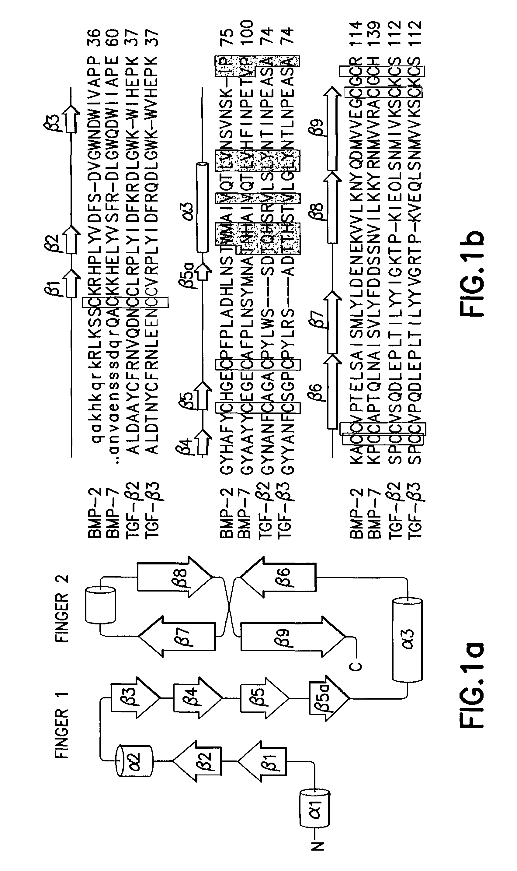
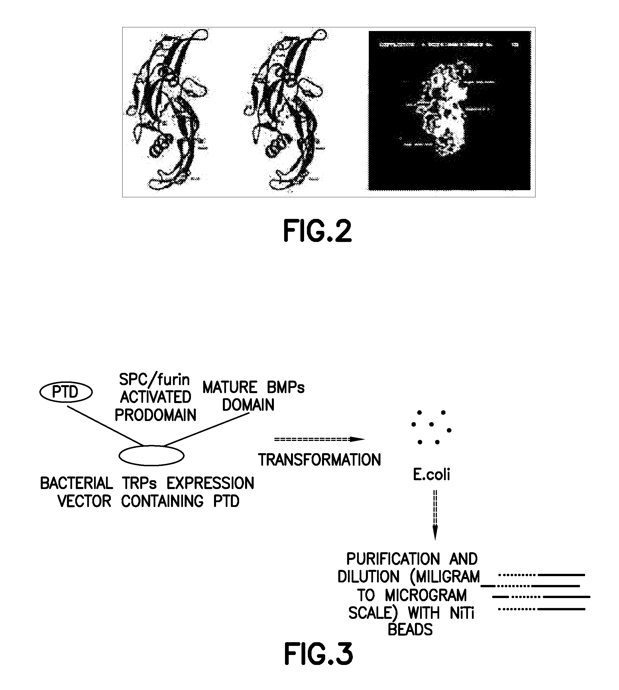
[0088]Each of proteins belonging to a BMP / TGF-β group consists of a dimer comprising the same two peptides linked to each other by one disulfide bond. In this regard, each of the peptides consists of 120-140 amino acids depending on the type of BMPs, one of 7 cystein terminal groups present in BMPs forms a disulfide bond with the same site of the other peptides to form a dimer, and the remaining 6 cysteins form 3 intrachain disulfide bonds in the same amino acids, resulting in a unique three-dimensional structure (Proc. Natl. Acad. Sci., 93:878, 1996; J. Bone Joint Surg., 83:S1, 2001). Herein, BMP2 (SEQ ID NO: 1) consists of 114 amino acids, BMP7 (SEQ ID NO: 40) consists of 139 amino acids, and each of TGF-β2 (SEQ ID NO: 41) and TGF-β3 (SEQ ID NO: 42) consists of 112 amino acids. FIG. 1 shows the amino acid sequences of these peptides and a schematic diagram of the three-dimensional structure thereof, and FIG. 2 shows the three-dimensiona...
example 2
Preparation of [TAT-FAD-hBMP2] Fusion Polypeptide (TRP-1)
[0099]As described in Example 1, hBMP2 shows biological activity by forming a characteristic internal three-dimensional structure and a dimer by a disulfide bond. However, as described in Example 1, a fusion protein of TAT and hBMP2 had no biological activity to induce bone cell differentiation even though it successfully permeated the cell membrane. This means that proteins having biological activity by secretion are insufficient to show activity only with the restructuring of amino acids by, e.g., HSPs, unlike the prior PTD-fused proteins (Trends Cell Biol., 10:290, 2000; Curr. Prot. Pept. Sci., 4:97, 2003).
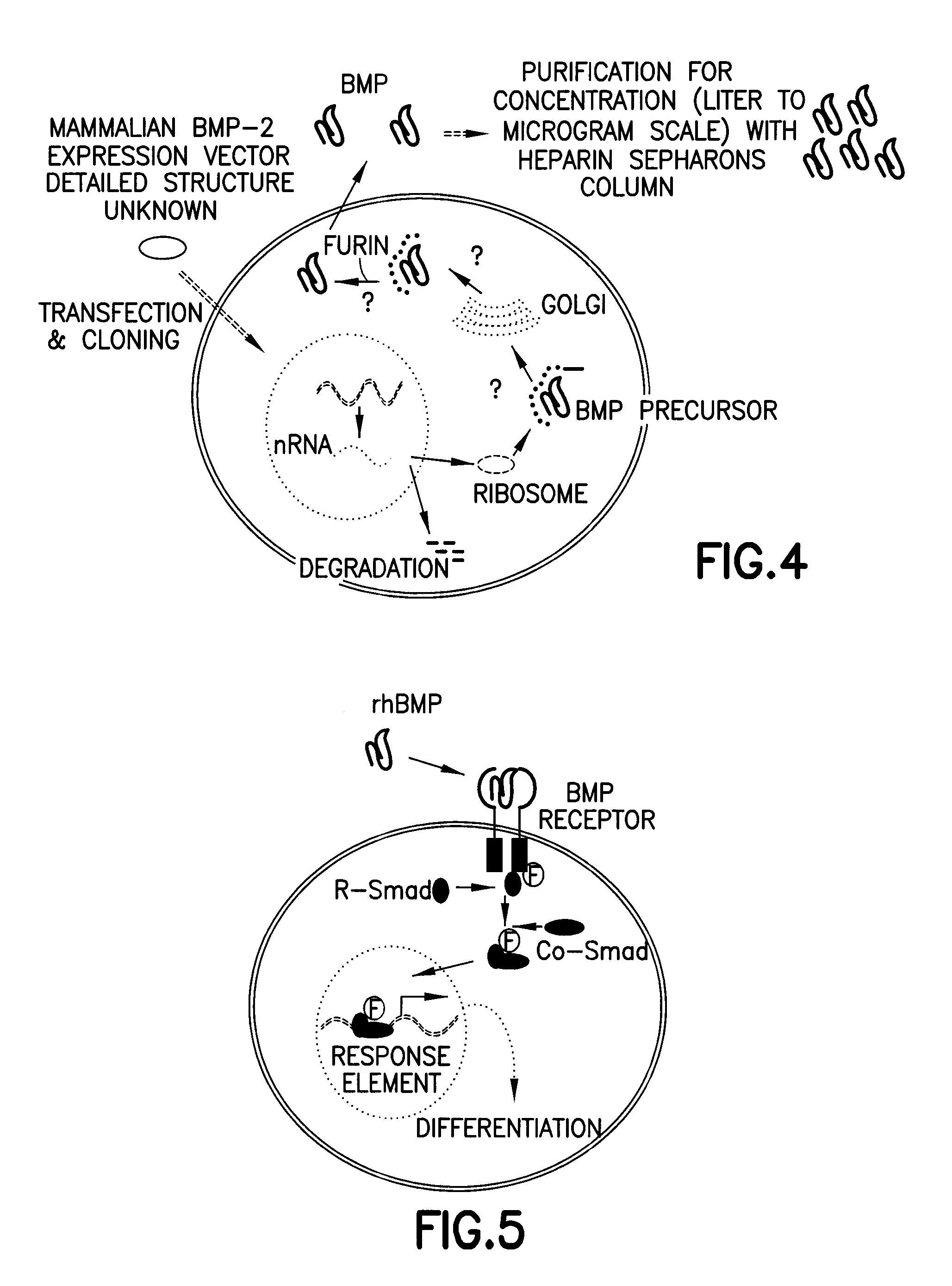
[0100]BMPs and TGF-β are present in a form of precursor consisting of more than 400 amino acids, when biosynthesized in intracellular ribosomes. The synthesized amino acids transport to Golgi complexes, endosomes, etc., by signal peptides present in the N-terminal region, and cleaved and activated by proprotein convertase...
example 3
Cleavage and Activation of TRP-1 by Proprotein Convertase
[0110]In order to examine whether the inventive non-activated TRP-1 is cleaved and activated by furin in cells, TRP-1 prepared in Example 2 was cleaved in vitro using a recombinant furin protein (Sigma, USA). As a result, as shown in FIG. 15, TRP-1 was successfully cleaved in vitro by the furin.
[0111]Also, in order to examine whether the inventive non-activated TRP-1 is activated by furin in cells, intracellular furin was inhibited using α1 antitrypsin Portland (α1-PDX) expression vector (Proc. Natl. Acad. Sci., 95:7293, 1998), as a furin inhibitory protein. Because the inventive non-activated TRP-1 is converted to a biochemically active protein after introduction into cells, the amount of TRP-1 initially introduced into cells shall gradually decrease at a given time after the administration of TRP-1. Thus, it can be expected that, when furin is inhibited by inducing the expression of α1-PDX, the remaining amount of TRP shall ...
PUM
| Property | Measurement | Unit |
|---|---|---|
| molecular weight | aaaaa | aaaaa |
| molecular weight | aaaaa | aaaaa |
| half life | aaaaa | aaaaa |
Abstract
Description
Claims
Application Information