Non-activated polypeptides having a function of tissue regeneration and method for preparing the same

Inactive Publication Date: 2007-05-10
KIM JUNG MOON +3
View PDF20 Cites 3 Cited by
  • Summary
  • Abstract
  • Description
  • Claims
  • Application Information

AI Technical Summary

Benefits of technology

[0025] The non-activated TRPs according to the present invention have no three-dimensional stereoregularity that are possessed in common by the previously known active BMPs. The TRPs are not biologically active by themselves yet before administered into patients. When the non-activated TRPs are administered to the human beings or mammals, however, the proprotein convertase cleavage sites of FAD in the TRPs are cleaved by proprotein convertase present in living cells, whereby TRD is activated, and the activated TRD is secreted out of the cells, thereby showing the desired tissue regeneration potencies. The TRPs according to the present invention are preferably in the form of a fusion polypeptide of PTD, FAD and TRD. The inventive TRPs have a function to stimulate the formation or regeneration of tissues, such as human bones and cartilages, or to improve the fibrosis or cirrhosis of organs, such as kidneys, liver, lungs and heart, and ultimately to

Problems solved by technology

As an old method for preparing BMPs, a method of extracting BMPs from demineralized animal bone tissue using natural salt (U.S. Pat. No. 4,294,753) has been attempted, but the method has problems in that the preparation efficiency is too low for mass production.
However, the methods for preparing recombinant hBMP2 and hBMP7 using the transformed CHO cells have problems in that the culturing of a large amount of CHO cells is required to obtain enough amount of active rhBMP2 or rhBMP7.
The separation and purification steps are very complicated, and the production cost is very high.
Also, these methods have a common problem that the biological activity of the proteins is reduced during the separation, purification, storage, medication and/or administration processes.
In this method of preparing the rhBMP14, however, the step

Method used


Image

  • Non-activated polypeptides having a function of tissue regeneration and method for preparing the same
  • Non-activated polypeptides having a function of tissue regeneration and method for preparing the same
  • Non-activated polypeptides having a function of tissue regeneration and method for preparing the same

Examples


Example

[0092] Example 1: Preparation of ITAT-hBMP2] fusion polypeptide

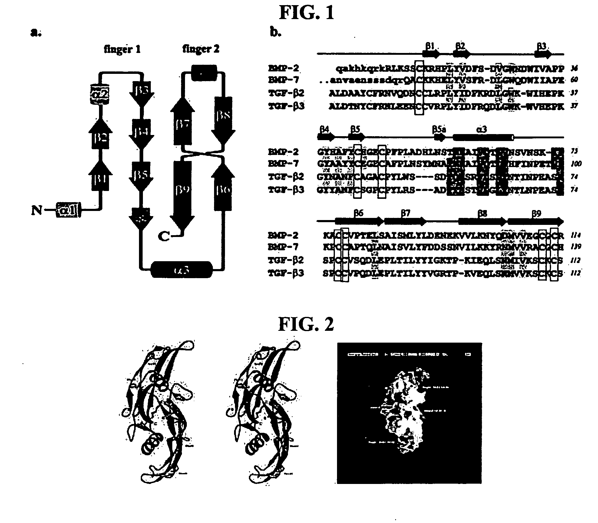
[0093] Each of proteins belonging to a BMP / TGF-β group consists of a dimer comprising the same two peptides linked to each other by one disulfide bond. In this regard, each of the peptides consists of 120-140 amino acids depending on the type of BMPs, one of 7 cystein terminal groups present in BMPs forms a disulfide bond with the same site of the other peptides to form a dimer, and the remaining 6 cysteins form 3 intrachain disulfide bonds in the same amino acids, resulting in a unique three-dimensional structure (Proc. Natl. Acad. Sci., 93:878, 1996; J Bone Joint Surg., 83:S1, 2001). Herein, BMP2 consists of 114 amino acids, BMP7 consists of 139 amino acids, and each of TGF-β and β3 consists of 112 amino acids. FIG. 1 shows the amino acid sequences of these peptides and a schematic diagram of the three-dimensional structure thereof, and FIG. 2 shows the three-dimensional structure of previously known BMP2 (Eur. J Bioc...

Example

[0103] Example 2: Preparation of ITAT-FAD-hBMP21 fusion polypeptide (TRP-1)

[0104] As described in Example 1, hBMP2 shows biological activity by forming a characteristic internal three-dimensional structure and a dimer by a disulfide bond. However, as described in Example 1, a fusion protein of TAT and hBMP2 had no biological activity to induce bone cell differentiation even though it successfully permeated the cell membrane. This means that proteins having biological activity by secretion are insufficient to show activity only with the restructuring of amino acids by, e.g., HSPs, unlike the prior PTD-fused proteins (Trends Cell BioL, 10:290, 2000; Curr. Prot. Pept. Sci., 4:97, 2003).

[0105] BMPs and TGF-β are present in a form of precursor consisting of more than 400 amino acids, when biosynthesized in intracellular ribosomes. The synthesized amino acids transport to Golgi complexes, endosomes, etc., by signal peptides present in the N-terminal region, and cleaved and activated by ...

Example

[0113] Example 3: Cleavage and activation of TRP-1 bv proprotein convertase

[0114] In order to examine whether the inventive non-activated TRP-1 is cleaved and activated by furin in cells, TRP- 1 prepared in Example 2 was cleaved in vitro using a recombinant furin protein (Sigma, USA). As a result, as shown in FIG. 15, TRP-1 was successfully cleaved in vitro by the furin.

[0115] Also, in order to examine whether the inventive non-activated TRP- 1 is activated by furin in cells, intracellular furin was inhibited using aL antitrypsin Portland (α1-PDX) expression vector (Proc. Natl. Acad. Sci., 95:7293, 1998), as a furin inhibitory protein. Because the inventive non-activated TRP-1 is converted to a biochemically active protein after introduction into cells, the amount of TRP- 1 initially introduced into cells shall gradually decrease at a given time after the administration of TRP-1. Thus, it can be expected that, when furin is inhibited by inducing the expression of α1-PDX, the remai...


PUM

PropertyMeasurementUnit
Compositionaaaaaaaaaa
Biological propertiesaaaaaaaaaa

Abstract

Non-activated tissue-regeneration polypeptides (TRPs) and the preparation methods thereof are disclosed. The TRPs contain: a protein transduction domain (PTD) making the polypeptides to permeate a cell membrane without cell membrane receptors; a furin activation domain (FAD) which has at least one proprotein convertase cleavage site and which can be cleaved by the proprotein convertase and activate a tissue regeneration domain (TRD) in cells; and a tissue regeneration domain (TRD) which can be activated by the proprotein convertase cleavage of the FAD to stimulate the growth or formation of tissues or to induce the regeneration of tissues. The TRPs can be practically mass-produced by the culture of bacteria, such as recombinant E. coli, and are in a non-activated state before in vivo administration. Thus, their production cost is only a few tenths of the prior active proteins having uses similar thereto, and processes for their separation, purification, handling, storage and administration are significantly simple and convenient. The in vivo administration of the TRPs can stimulate the formation or regeneration of tissues, such as bones or cartilages, or improve the fibrosis and cirrhosis of organs, such as kidneys, liver, lungs and heart by pharmacological mechanisims completely different from those of prior rhBMPs or TGF-β proteins. Accordingly, the TRPs will be useful as drugs having new mechanisms.

Description

TECHNICAL FIELD [0001] The present invention is related to non-activated tissue-regenerative polypeptides (TRPs) and the methods for preparing the TRPs, the tissue regenerative polypeptides, which contain: a protein transduction domain (PTD) making the polypeptides to permeate a cell membrane without cell membrane receptors; a furin activation domain (FAD) which has at least one proprotein convertase cleavage site and which can be cleaved by the proprotein convertase and can activate a non-activated tissue regeneration domain (TRD) in cells; and a non-activated tissue regeneration domain (TRD) which can be activated through the the cleavage of the FAD by the proprotein convertase in cells and which can stimulate the growth or formation of tissues or to induce tissue regeneration. BACKGROUND ART [0002] It is known that bone morphogenetic proteins (BMPs) stimulate the healing of bone defects in mammals or Primates, and particularly are secreted in bone cells to induce bone formation t...

Claims


Application Information

Patent Timeline
IPC IPC(8): A61K38/17C07K14/475C07H21/04C12P21/06
CPCC07K14/51
Inventor KIM, JUNG MOONKIM, TAE HANLEE, JONG SUKYOOK, JONG IN
Owner KIM JUNG MOON
Why Eureka
Try Eureka