
Methods of making frozen tissue microarrays

a tissue array and microarray technology, applied in the field of frozen tissue arrays, can solve the problems of slow and tedious process, h&e can reveal only h&e can only reveal a limited amount of diagnostic information,

Inactive Publication Date: 2012-10-02
NUCLEA BIOMARKERS
View PDF23 Cites 26 Cited by
  • Summary
  • Abstract
  • Description
  • Claims
  • Application Information

AI Technical Summary

Benefits of technology

"The invention provides microarrays made from frozen tissue and cell samples that can be used for high throughput analysis of nucleic acids and proteins. These microarrays can be prepared by sectioning a block of frozen embedding material and filling the sections with the donor samples. The donor samples can be obtained from a variety of sources such as humans, animals, plants, and non-human animals. The microarrays can be used to evaluate the expression of genes and proteins in different tissues and cells, and can also be used to identify diagnostic markers for diseases. The invention also provides methods for preparing and using the microarrays."

Problems solved by technology

However, standard staining methods such as hematoxylin-eosin (H&E) generally can reveal only a limited amount of diagnostic information.
However, the development of new molecular markers of clinical importance has been impeded by the slow and tedious process of determining the expression of these markers in large numbers of clinical specimens.
As the number of molecular probes increases, the number of tissue samples which can be evaluated in a single experiment becomes a rate-limiting factor.
In addition to requiring a high degree of manual dexterity, these methods randomly arranged samples, making it difficult to identify specimens of interest.
102: 192–193, however, these methods are also labor intensive and time consuming.
While greatly increasing the throughput of methods involving the use of tissue microarrays, the technique has been limited to the evaluation of paraffin-embedded tissue specimens which are not optimal for many types of molecular analyses.

Method used


Image

  • Methods of making frozen tissue microarrays
  • Methods of making frozen tissue microarrays
  • Methods of making frozen tissue microarrays

Examples


Embodiment Construction

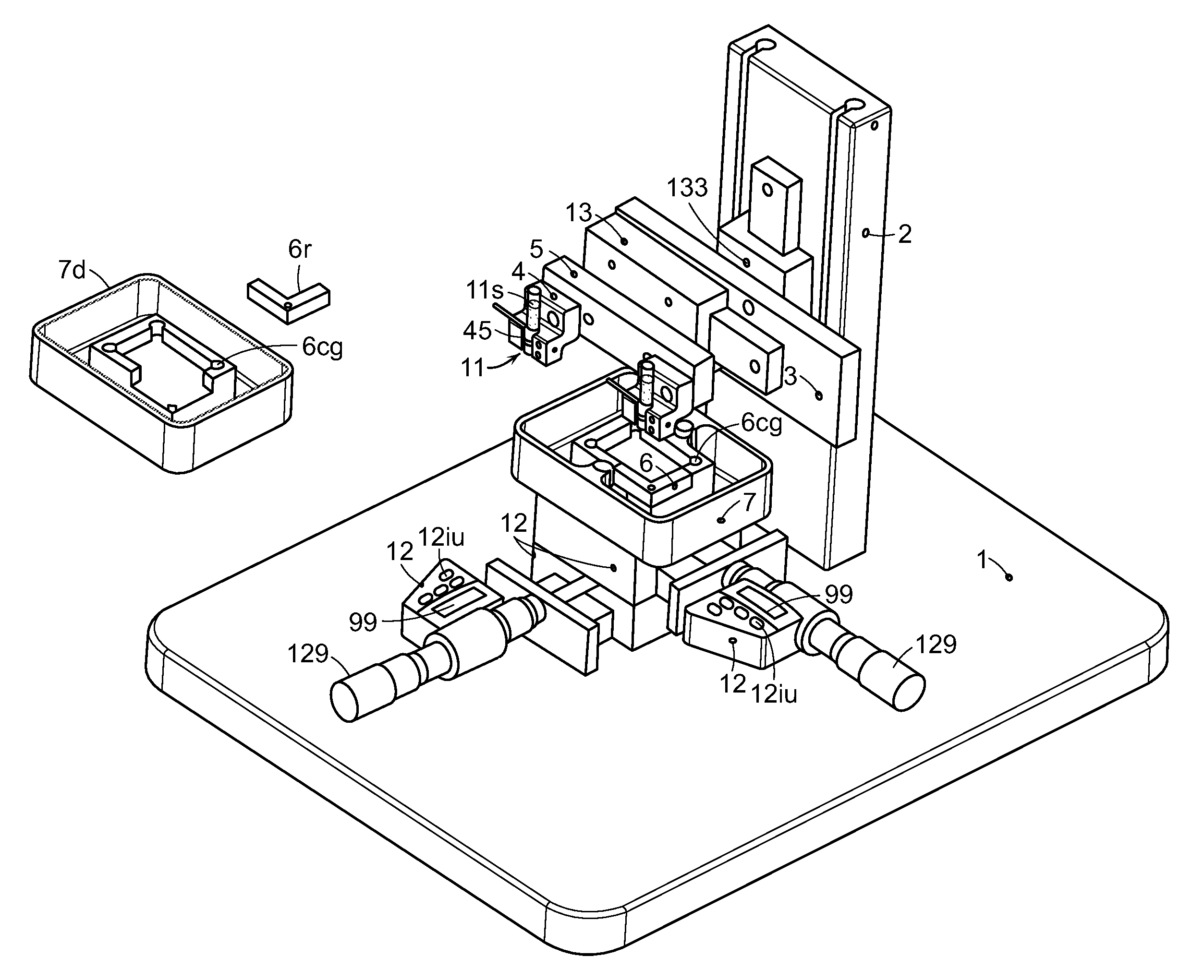
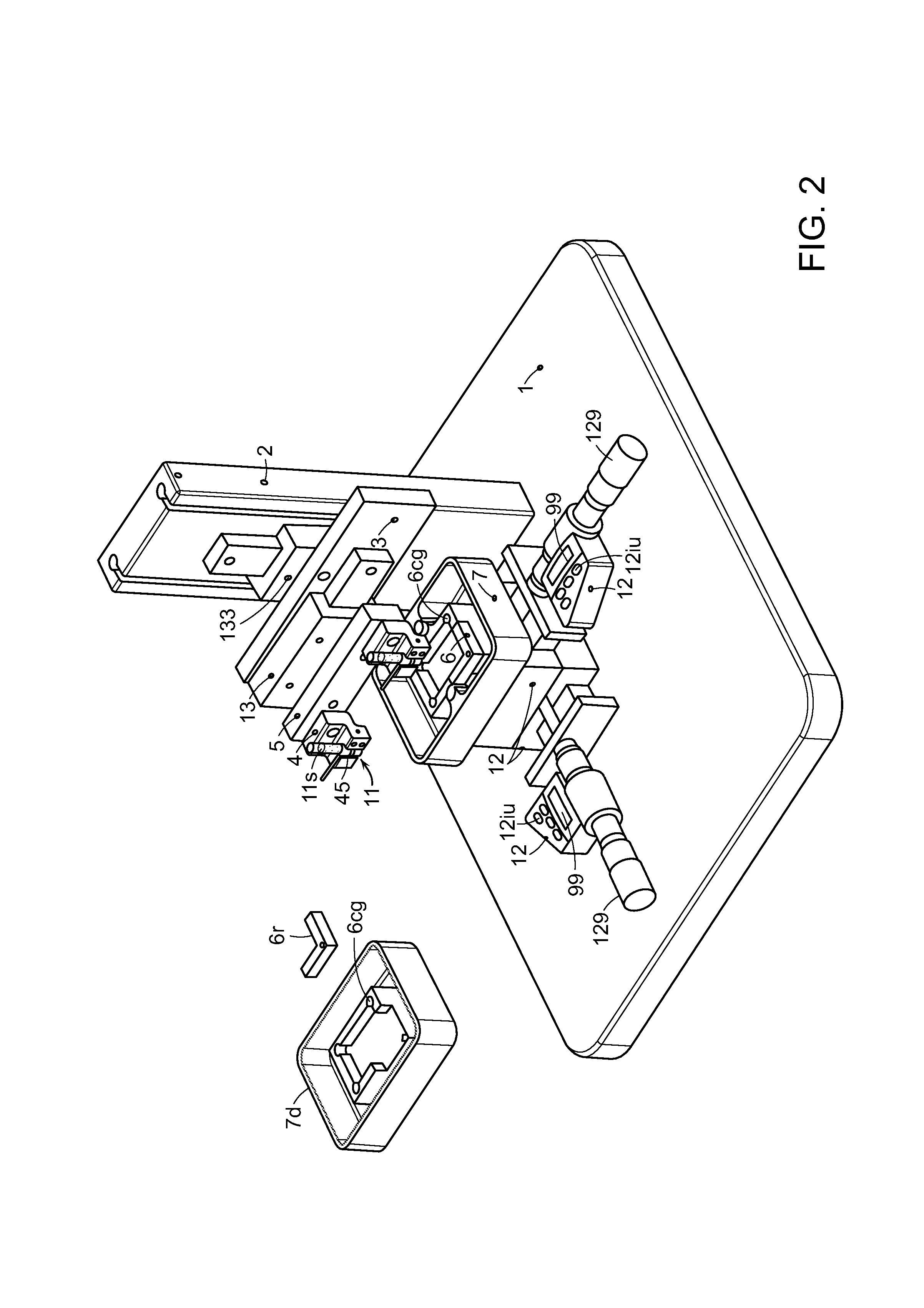
[0025]The invention provides a plurality of frozen tissue samples embedded in a single microarray block for generating hundreds of substantially identical frozen tissue microarrays and a precision instrument for generating the same. Frozen tissue microarrays according to the invention can be evaluated in high throughput parallel analyses using the same or different molecular probes, enabling gene identification, molecular profiling, selection of promising drug targets, sorting and prioritizing of expressed sequence array data, and the identification of abnormal physiological processes associated with disease.

[0026]Definitions

[0027]The following definitions are provided for specific terms which are used in the following written description.

[0028]The term “frozen” as used herein, refers to temperatures which are at least −20° C. or colder.

[0029]As used herein “donor block” refers to a fast-freezing embedding material comprising a frozen tissue or cell(s). While referred to as a “block...


PUM

PropertyMeasurementUnit
diameteraaaaaaaaaa
diameteraaaaaaaaaa
sizesaaaaaaaaaa

Abstract

The invention provides microarrays comprising a plurality of frozen tissues and / or cell samples and methods of preparing and using the same. By using frozen samples, the microarrays provide optimal samples from which to detect the expression of both nucleic acids (e.g., mRNAs) and proteins in high throughput parallel analyses. The microarrays enable gene identification, molecular profiling, selection of promising drug targets, sorting and prioritizing of expressed sequence array data, and the identification of abnormal physiological processes associated with disease.

Description

RELATED APPLICATIONS [0001]This application claims priority under 35 U.S.C. § 119(e) to U.S. Provisional Application Ser. No. 60 / 213,321, filed Jun. 22, 2000, U.S. Provisional Application No. 60 / 234,493, filed Sep. 22, 2000, and U.S. Provisional Application No. 60 / 236,649, filed Sep. 29, 2000, the entireties of which are incorporated by reference herein.FIELD OF THE INVENTION [0002]The present invention relates to frozen tissue arrays and methods of arraying frozen tissue samples for high throughput molecular analyses.BACKGROUND OF THE INVENTION [0003]The microscopic examination and classification of tissues has improved medical treatment. For example, in the case of many tumors, a diagnosis can be made on the basis of cell morphology and staining characteristics. Even the aggressiveness of a tumor can sometimes be predicted through microscopic evaluation. However, standard staining methods such as hematoxylin-eosin (H&E) generally can reveal only a limited amount of diagnostic info...

Claims


Application Information

Patent Timeline
Patent Type & Authority Patents(United States)
IPC IPC(8): C12Q1/68C12N11/16G01N15/06B01J19/00G01N33/53C12M1/00C12N15/09C40B30/04G01N33/574G01N33/68G01N37/00
CPCB01J19/0046C12Q1/6837C12Q1/6886C40B30/04G01N33/574G01N33/6845B01J2219/00274B01J2219/00673B01J2219/00743C12Q1/6841C12Q1/6883C12Q2565/513
Inventor MURACA, PATRICK J.
Owner NUCLEA BIOMARKERS
Learn More