Capsule endoscopic system and operation control method of capsule endoscope

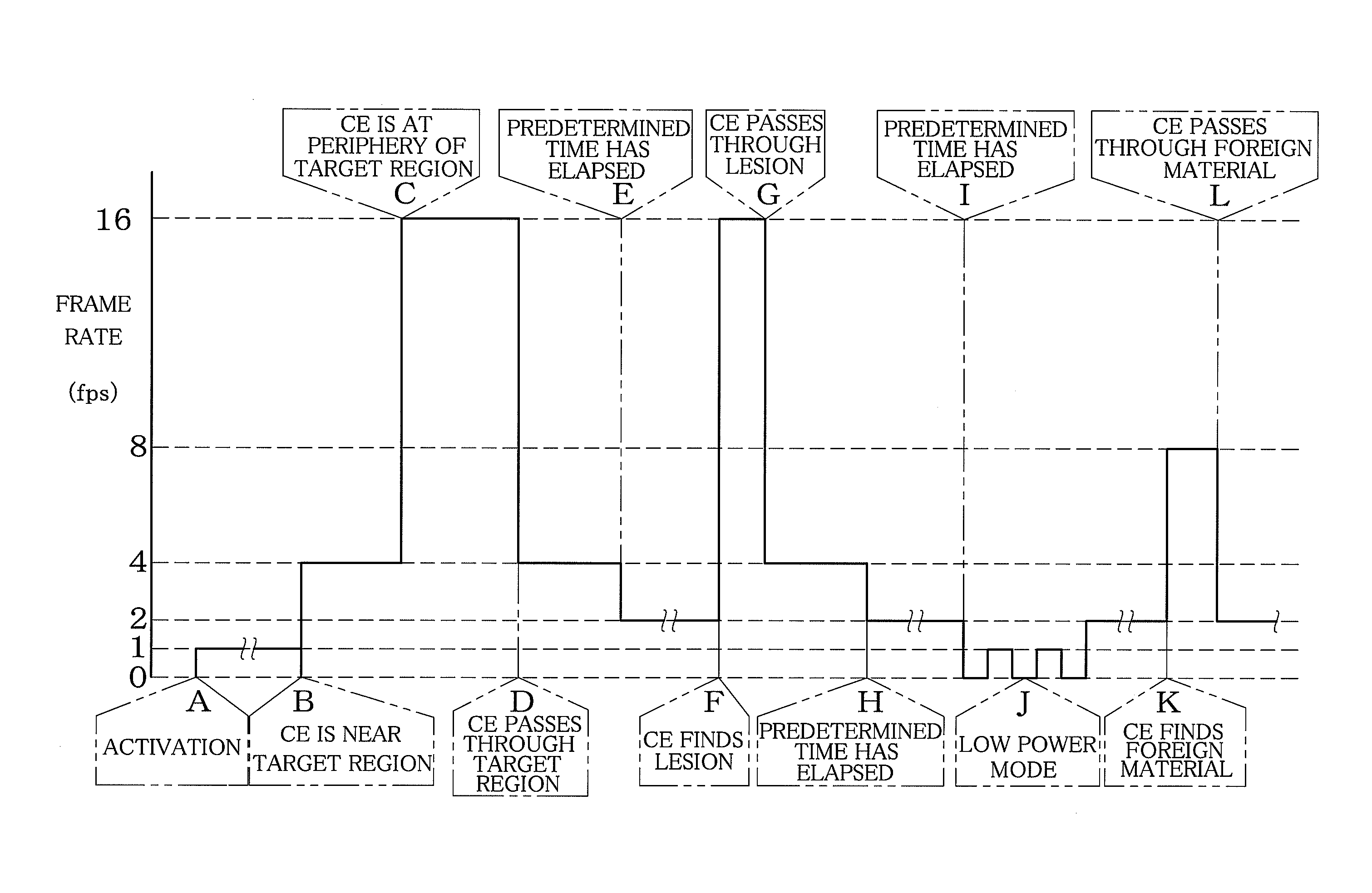
a capsule endoscope and capsule technology, applied in the field of capsule endoscopic system and operation control method of capsule endoscope, can solve the problems of not being able to read the target region in detail, taking a lot of time and work, and the image data stored in the receiver is huge, so as to achieve the effect of short time, reduced frame rate and increased frame ra

Inactive Publication Date: 2013-08-27
FUJIFILM CORP
View PDF12 Cites 0 Cited by
  • Summary
  • Abstract
  • Description
  • Claims
  • Application Information

AI Technical Summary

Benefits of technology

[0012]In view of the above, an object of the present invention is to provide a capsule endoscopic system capable of reading an image of a target region more in detail in a short time, and to provide an operation control method of this capsule endoscope.
[0029]Moreover, according to the present invention, there is provided an operation control method of capsule endoscope including the steps of : retrieving past diagnostic information of a patient to be inspected from an information management device to a receiver; comparing and analyzing the diagnostic information and present information obtained by a capsule endoscope during endoscopy; producing a control command to control operation of each component of the capsule endoscope based on an analysis result of the analyzing step; wirelessly transmitting and receiving the control command between the receiver and the capsule endoscope; and causing each component of the capsule endoscope to operate in accordance with the control command.
[0030]According to the capsule endoscopic system and the operation control method of the capsule endoscope of the present invention, based on the comparison result between the past diagnostic information and the present information obtained by the capsule endoscope during the endoscopy, the operation of each component of the capsule endoscope is controlled. Therefore, if the image of the target region and the position information are prepared as the diagnostic information, the target region can be identified in comparison with the present information. This enables controlling the capsule endoscope such that, for example, when the capsule endoscope is passing through the target region, the frame rate can be raised, and after the capsule endoscope has passed through the target region, the frame rate can be lowered. As a result, it is possible to reading images of the target region more in detail in a short time.

Problems solved by technology

The image shooting is performed for at least eight hours, and the amount of image data stored in the receiver is huge.
Therefore, in a case where the image is read out for diagnosis after the endoscopy, when all the shot images are to be read out, it takes a lot of time and work.
Therefore, there is a fear that it becomes impossible to read an image of the target region in detail.
Therefore, as in the case of the Japanese Patent Translation Publication No. 2004-521662, the Japanese Patent Application Laid-open Publication No. 2006-223892, and the Japanese Patent Application Laid-open Publication No. 2006-288808, the demand for reading images of the target region more in detail in a short time can not be achieved.

Method used


Image

  • Capsule endoscopic system and operation control method of capsule endoscope
  • Capsule endoscopic system and operation control method of capsule endoscope
  • Capsule endoscopic system and operation control method of capsule endoscope

Examples


Embodiment Construction

[0051]Embodiments of the present invention are described hereinbelow. The present invention, however, is not limited to the following embodiments.

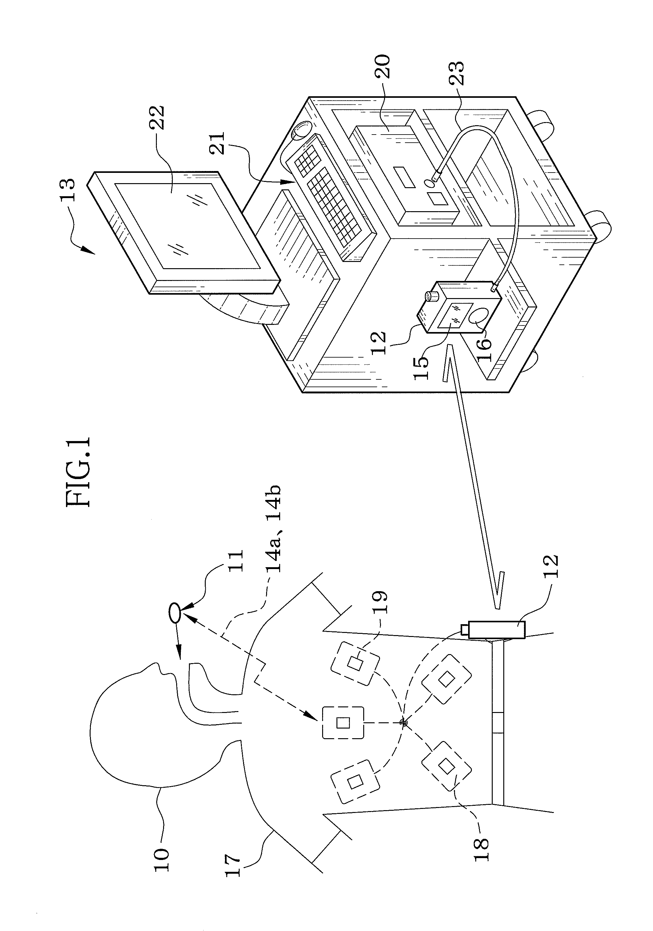
[0052]As shown in FIG. 1, a capsule endoscopic system includes a capsule endoscope (hereinafter abbreviated as CE) 11, a receiver 12, and a work station (hereinafter abbreviated as WS) 13. The capsule endoscope 11 is taken into a body through a mouth of a patient 10. The receiver 12 is attached to a belt of the patient 10. A doctor uses the WS 13 to read an image obtained by the CE 11 for diagnosis.

[0053]While passing through tracts in the human body, the CE 11 shoots images of inner wall surface of the tracts. The obtained image data is wirelessly transmitted to the receiver 12 via a radio wave 14a. Further, the CE 11 wirelessly receives a control command from the receiver 12 via a radio wave 14b, and operates in accordance with the control command.

[0054]The receiver 12 includes a liquid crystal display (hereinafter abbreviated as LCD) 15...


PUM


Abstract

A receiver of capsule endoscopic system downloads diagnostic information of a patient obtained by a past diagnosis from a data storage of work station. A data analyzer of the receiver compares the diagnostic information and present information obtained by use of a capsule endoscope during endoscopy. A CPU produces a control command in which a frame rate of image shooting by use of the capsule endoscope is set based on an analysis result of the data analyzer. The produced control command is wirelessly transmitted via a radio wave from the receiver. The capsule endoscope wirelessly receives the control command from the receiver via the radio wave, and performs image shooting with the frame rate set by the control command.

Description

FIELD OF THE INVENTION[0001]The present invention relates to a capsule endoscopic system in which an image inside a body of a patient obtained by a capsule endoscope is used for medical diagnosis, and an operation control method of the capsule endoscope.BACKGROUND OF THE INVENTION[0002]Recently, medical diagnosis by use of a capsule endoscope incorporating an image sensor, a light source, and the like in an extremely small capsule has been put to practical use. In the medical diagnosis by use of the capsule endoscope, at first, the capsule endoscope is swallowed by a patient, and while a region to be observed inside a body of a patient (inner wall surface of tracts in the human body) is illuminated by the light source, the region to be observed is shot by the image sensor. Image data thus obtained is wirelessly transmitted to a receiver, and sequentially stored in a recording medium such as a flash memory provided in the receiver. During or after the endoscopy, the image data is ret...

Claims


Application Information

Patent Timeline
Patent Type & Authority Patents(United States)
IPC IPC(8): A61B1/00A61B1/04A61B1/06
CPCA61B1/00016A61B1/00036A61B1/041A61B1/045A61B1/0638A61B5/065A61B5/14539A61B2560/0209A61B2562/0219
Inventor NISHINO, NAOYUKI
Owner FUJIFILM CORP
Why Eureka
Try Eureka