System for performing and monitoring minimally invasive interventions

a technology for performing and monitoring systems, applied in the field of systems for performing and monitoring minimally invasive interventions, can solve the problem that mobile devices do not as a rule achieve the level of x-ray performan

Inactive Publication Date: 2013-10-01
SIEMENS HEALTHCARE GMBH
View PDF36 Cites 45 Cited by
  • Summary
  • Abstract
  • Description
  • Claims
  • Application Information

AI Technical Summary

Benefits of technology

[0009]The present system allows all the steps needed for treating tachycardial arrhythmias to be carried out in an electrophysiological laboratory without requiring further imaging resources. All tachycardial arrhythmias can thus be eliminated reliably, at little risk to the patient and to the clinical staff, with a high level of quality and with good therapeutic success. In this situation, the system is not dependent on preliminary images from computer tomographs or magnetic resonance tomographs. Rather, the present system allows 3D image data to be generated in real time and for example to have 2D images superimposed on it. With the existing system, the 3D images are captured by the X-ray unit configured for this purpose, which allows a 3D image data set to be reconstructed from different selectable projections. The techniques for the reconstruction of a 3D image data set from images taken by the X-ray unit employed in the present system, in particular a C-arm X-ray unit, are known as a general principle. 3D images of a skull and the vessels, for example, can thus be obtained by using the AXIOM Artis® FA / FB equipment from Siemens with an associated workstation. A method for generating a volume date set is likewise known from US 2004 / 0066906 A1. Further examples of C-arm X-ray units which deliver 3D images are described in Electromedica 70 (2002) No. 1, “Initial Clinical Experiences with the SIREMOBIL ISO-C3D” by Euler et al. on pages 48 to 51, in DE 100 47 364 A1, in U.S. Pat. No. 6,379,041 or in DE 103 06 068 A1. The majority of previously known solutions however utilize a mobile C-arm X-ray unit for generating the 3D X-ray images. Such types of mobile devices do not however as a rule achieve levels of X-ray performance which are sufficient for cardiological images. A stationary X-ray unit, for example an AXIOM Artis® FC, TC or BC with a flat detector, is therefore to be preferred for the present system.
[0011]A further significant advantage of the present system consists in the capability for data exchange between all the connected units. In this situation the operator does not need to transfer any data or information whatsoever from one unit to the other unit in question, or enter any data or information there. Rather, the central control and evaluation unit ensures at all times that all the connected units have at their disposal at all times the data required for their use from the other unit or units. In particular, the present system can be operated in the preferred embodiment by way of a central operator interface at which all the required information and data is available. In this embodiment, at least one central screen is also provided, on which all the data generated by the different units or catheters is displayed, superimposed if need be.

Problems solved by technology

Such types of mobile devices do not however as a rule achieve levels of X-ray performance which are sufficient for cardiological images.

Method used


Image

  • System for performing and monitoring minimally invasive interventions
  • System for performing and monitoring minimally invasive interventions

Examples


Embodiment Construction

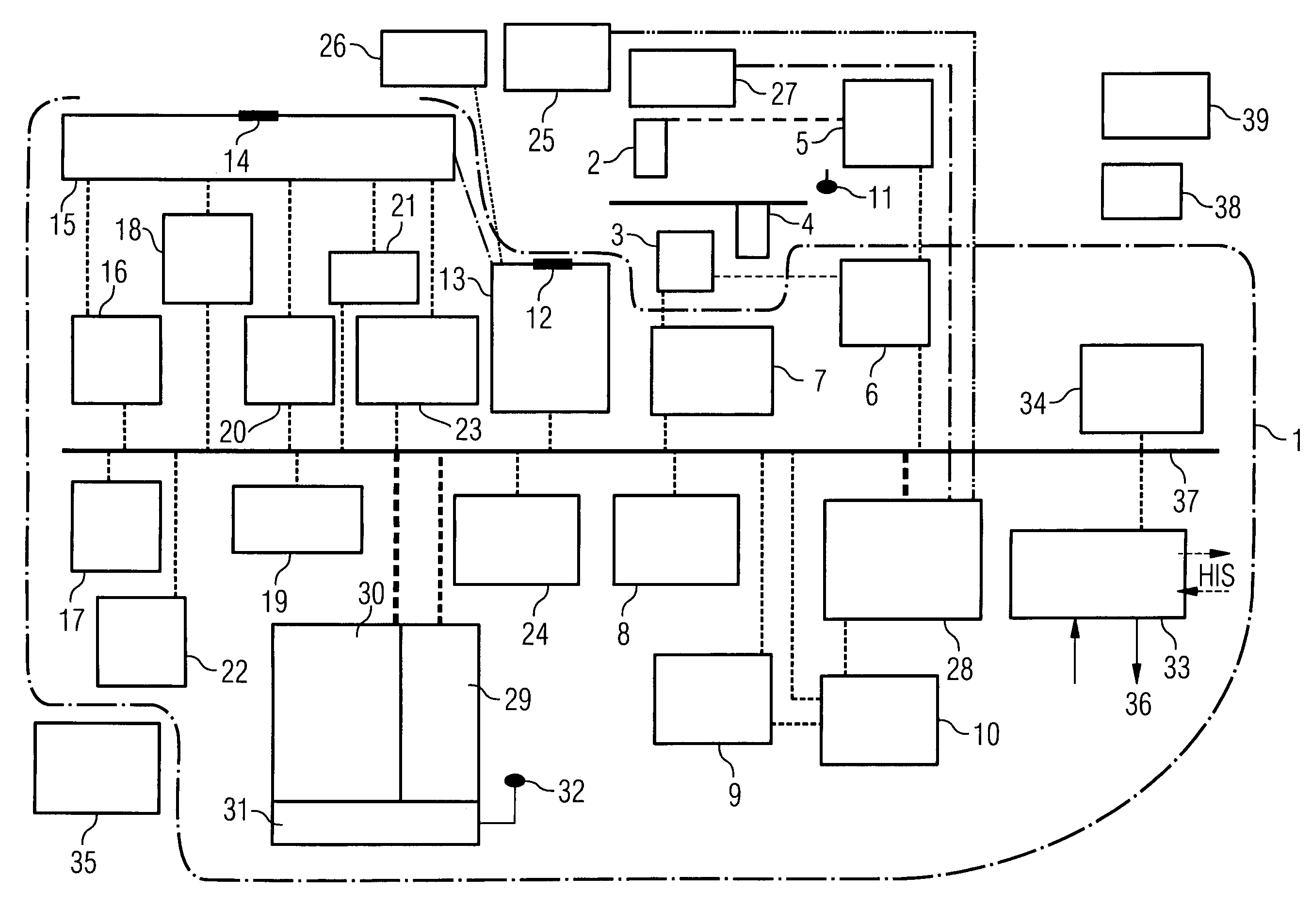
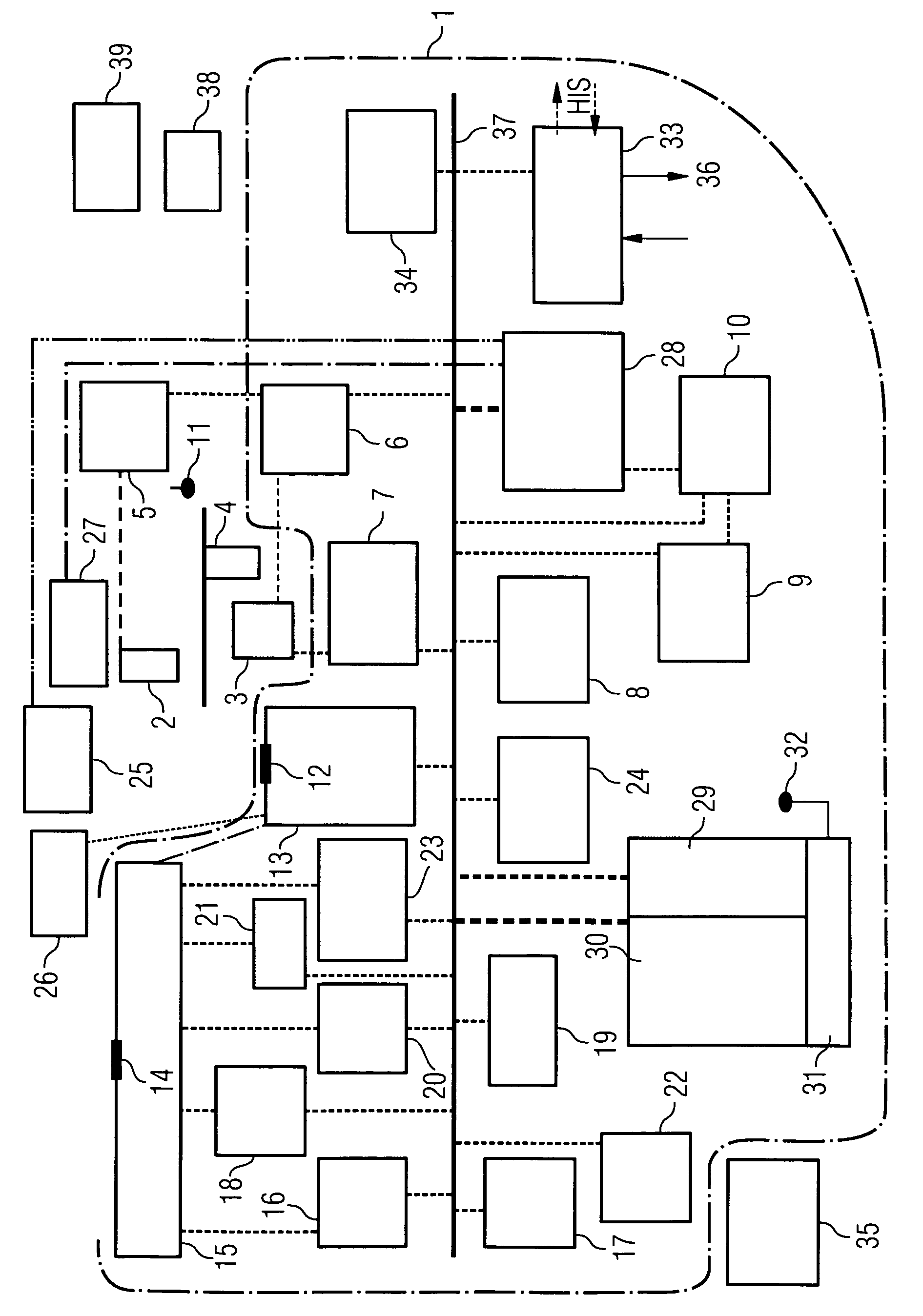
[0015]The attached FIGURE shows an illustration of the system in an embodiment which contains numerous optional components. The area enclosed within the dashed line here denotes the control and evaluation unit 1 with the associated modules. It is naturally also possible however for individual ones of these modules to be designed as part of the individual units, particularly if these modules perform a preprocessing of the captured measurement or image data which is as a rule required in the case of units or catheters of this kind.

[0016]The system illustrated in the FIGURE by way of example comprises an X-ray unit for cardiological examination which has at least one C-arm with an X-ray source 2, a radiation shutter and also an X-ray detector 3, for example with a flat detector or aSi detector, and a patient positioning table 4. In this situation the patient positioning table 4 can have an X-ray transparent surface for patient positioning. In the preferred embodiment, this patient posi...


PUM

PropertyMeasurementUnit
angleaaaaaaaaaa
angleaaaaaaaaaa
angleaaaaaaaaaa

Abstract

The present invention relates to a system for performing and monitoring minimally invasive interventions with an x-ray unit, in which at least one x-ray source and one x-ray detector can traverse a circular track through an angle range, an ECG recording unit, an imaging catheter, a mapping unit with a mapping catheter and an ablation unit with an ablation catheter. The system comprises a control and evaluation unit with interfaces for the units and catheters, which enable an exchange of data with the control and evaluation unit. The control and evaluation unit is designed for processing measurement or image data which it receives from the catheters and units, and for controlling the catheters and units for the capture of the measurement or image data. The workflow from the examination through to the therapy, particularly with regard to the treatment of tachycardial arrhythmias, is covered completely and continuously by the proposed system.

Description

CROSS REFERENCE TO RELATED APPLICATIONS[0001]This application claims priority of German application No. 10 2005 032 755.9 filed Jul. 13, 2005, which is incorporated by reference herein in its entirety.FIELD OF THE INVENTION[0002]The present invention relates to a system for performing and monitoring minimally invasive interventions, particularly for the treatment of electrophysiological diseases.BACKGROUND OF THE INVENTION[0003]The serious cardiological diseases include the tachycardial arrhythmias, such as atrial fibrillation for example. In this situation, the vestibule is very frequently excited by conduction disturbances in the heart. In the case of other, for example ventricular, tachycardias the result is a complete contraction and thus an inadequate pumping performance by the heart. In the past, an attempt was made either to reduce the effect of the atrial fibrillation by means of a continuous intake of medicines or to eliminate the cause of the atrial fibrillation by means o...

Claims


Application Information

Patent Timeline
Patent Type & Authority Patents(United States)
IPC IPC(8): A61B5/05A61B8/12A61N1/00
CPCA61B5/0215A61B6/466A61B6/481A61B6/504A61B6/5247A61B8/12A61B8/4416A61B18/1492A61B19/5244A61B6/584A61B5/11A61B5/721A61B6/541A61B8/481A61B2017/00039A61B2018/00125A61B2018/00839A61B2019/5238A61B2019/5251A61B2019/5276A61B2019/5278A61B2019/5291A61B5/0402A61B2090/3782A61B34/20A61B2090/376A61B2034/2051A61B2090/378A61B2090/365A61B2562/17A61B5/318
Inventor MASCHKE, MICHAELKILLMANN, REINMAR
Owner SIEMENS HEALTHCARE GMBH
Why Eureka
Try Eureka