
Method for repair of articular cartilage defect and a device used therein

a technology of articular cartilage and repair method, which is applied in the direction of prosthesis, drug composition, biocide, etc., can solve the problems of articular cartilage defects, relative slow metabolism, and serious and painful impact or rubbing of two bones,

Active Publication Date: 2013-12-03
TAIPEI MEDICAL UNIV
View PDF3 Cites 0 Cited by
  • Summary
  • Abstract
  • Description
  • Claims
  • Application Information

AI Technical Summary

Benefits of technology

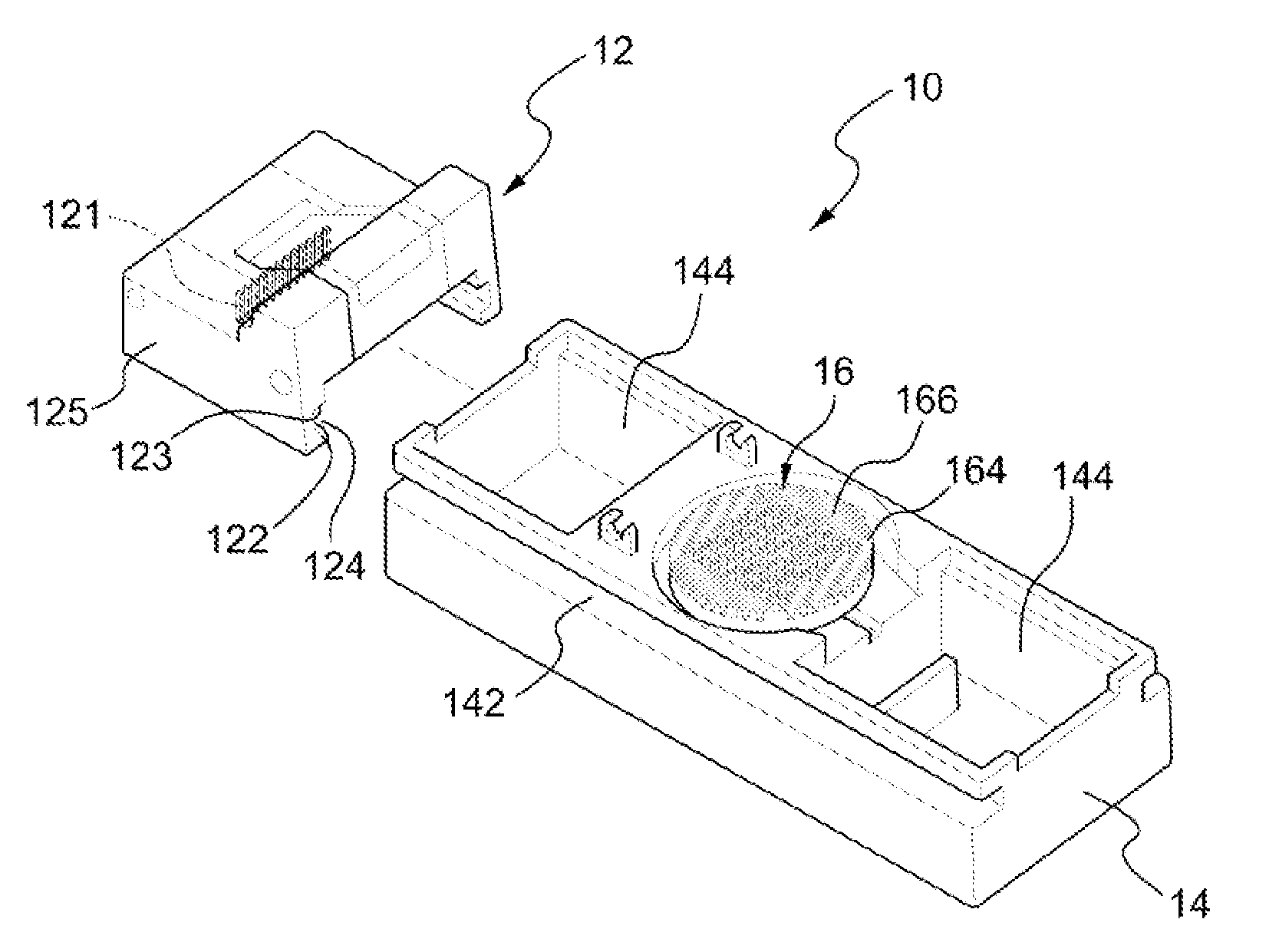
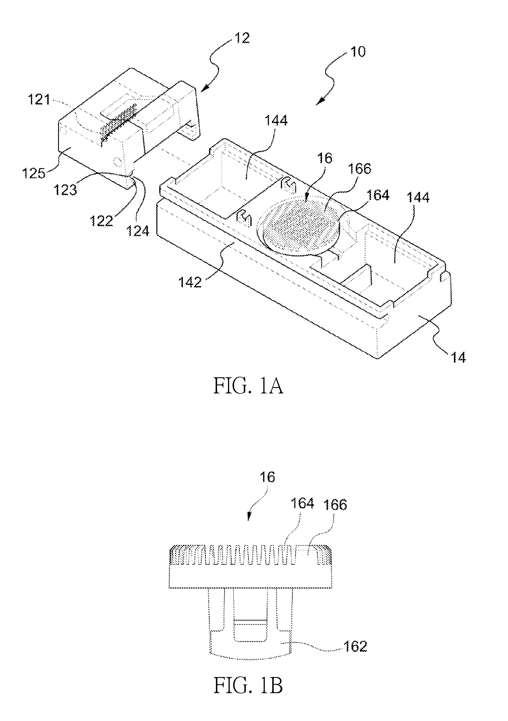
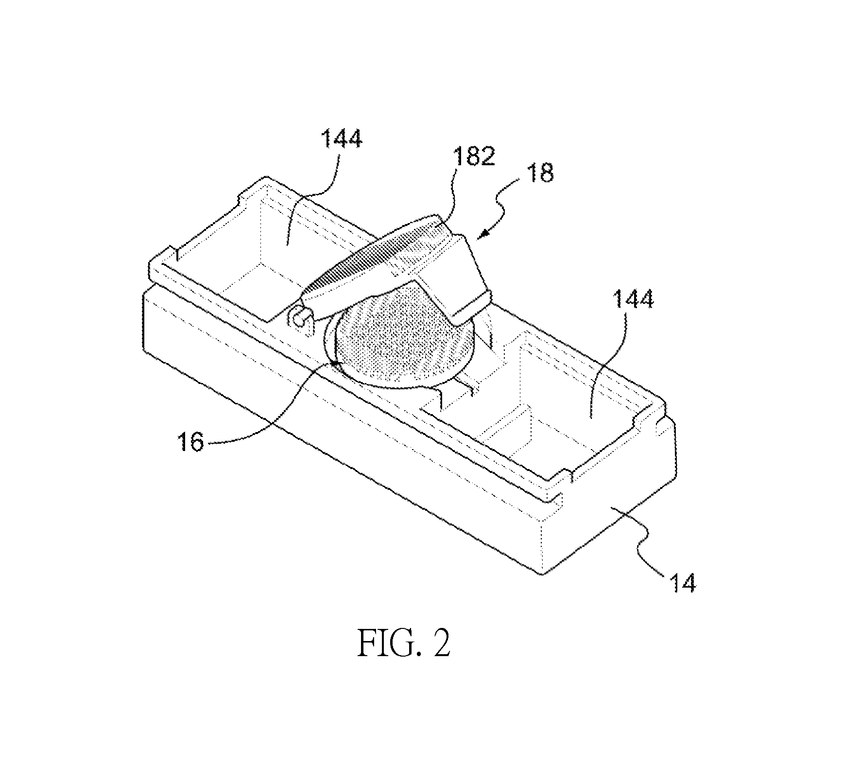
[0014]the blade holder having two parallel longitudinal sides, and an inwardly extending lower flange and an upper flange at each of the two parallel longitudinal sides of the blade holder; the lower flange and upper flange define a space to receive the slide rail, wherein said pair of slide rails slidably engages with said blade holder, allowing said blade holder to move in a longitudinal direction relative to said base, thereby permitting said plurality of blades to slice into the cartilage.

Problems solved by technology

Articular cartilage does not have any blood vessels or neurons, so its nutrients are absorbed from the surrounding joint fluids, and thus, nutrients are provided slowly, resulting in relatively slow metabolism.
Also, because a nerve system exists in bones, any impact or rubbing of two bones can be serious and painful.
Articular cartilage defects may be caused by wear over time, mechanical injuries or diseases.
Many arthritic diseases and different degrees of trauma can cause destruction or deterioration of this fragile layer, leading to pain, joint stiffness, and even crippling disability.
For example, joints are worn down over time; mechanical injuries are caused by accidents, causing the cartilage to tear and break down; and cartilage is eroded because of diseases, such as gout, rheumatoid arthritis and osteoarthritis.
It is generally believed that, because articular cartilage lacks a vasculature, damaged cartilage tissue does not receive sufficient or proper stimuli to elicit a repair response.
Once the articular cartilage becomes damaged, it cannot repair itself and therefore is worn down gradually.
In addition, if the bone underneath the articular cartilage becomes damaged, pain or swelling may occur, resulting in difficulty walking and affecting daily life.
However, this prior art reference did not teach nor suggest the optimal method for repair of articular cartilage defects.
Moreover, the xenograft and xenogenous biomaterials may cause immunological problems, and the aforementioned is not the method most suitable for solving problems with the repair and treatment of articular cartilage defects.
This manual labor-intense process is extremely costly and time consuming.
Although clinical data suggests long term benefits for the patient, the prohibitive cost of the procedure combined with the traumatic impact of two surgical procedures to the knee have hampered adoption of this technique.
Because the patient will have to endure the trauma of two surgical operations, the cartilage defects typically take a long time to repair.
Furthermore, cell culture in vitro has many disadvantages, such as contamination.

Method used


Image

  • Method for repair of articular cartilage defect and a device used therein
  • Method for repair of articular cartilage defect and a device used therein
  • Method for repair of articular cartilage defect and a device used therein

Examples


example

Example 1

Use of Cartilage Fragments in Combination with PRF in the Single Surgical Procedure

Preparation of PRF

[0046]Pigs are selected for use as laboratory animals in the present embodiment. Six ml blood is drawn from the pigs. The blood is collected in a container, such as a centrifuge tube, holding a separator polyester gel, and in the present embodiment, the centrifuge tube is used to contain the blood. The centrifuge tube containing the blood is centrifuged at a speed in the range of 1000 to 5000 rpm for 1 to 20 minutes, and preferably, also at 2500 to 3500 rpm for 8 to 12 minutes. The centrifuge time may be adjusted according to the centrifuge speed. After centrifugation, jelly-like PRF is obtained in the middle part of the centrifuge tube, and then sterile forceps are used to clip the jelly-like PRF out. All steps in the preparation of the PRF are performed under standard disinfection procedures. The 6 ml whole blood may yield 1-1.5 ml of PRF. The size of the autologous PRF is...


PUM


Abstract

The invention provides a single surgical procedure by using autologous platelet-rich fibrin (PRF) and cartilage fragments to solve problems of contamination during incubation of chondrocytes and transplant rejection in vivo, which has the benefit of a more efficient and shorter recovery time. The invention also provides a device for easily and rapidly cutting cartilage to cartilage fragments so as to reduce contamination of the implant and infection.

Description

CROSS-REFERENCE TO RELATED APPLICATIONS[0001]This continuation-in-part application claims priority to U.S. Non-Provisional patent application Ser. No. 12 / 825,287, filed Jun. 28, 2010, now abandoned, which claims priority to Taiwan Patent Application No. 098142566, filed Dec. 11, 2009. All of which are hereby incorporated by reference in their entirety.FIELD OF THE INVENTION[0002]The present invention relates to a method for the repair of articular cartilage defects. Particularly, the invention provides a single surgical procedure and a device used in the procedure.BACKGROUND OF THE INVENTION[0003]The healthy cartilage of joints (articular cartilage) in humans and other mammals is characterized by a columnar growth pattern of chondrocytes, which produce a hyaline cartilage predominantly containing proteoglycans, type II collagen, and water. Articular cartilage, which covers each end of the long bones in vivo, is a smooth white structure. The structure of the articular cartilage is si...

Claims


Application Information

Patent Timeline
Patent Type & Authority Patents(United States)
IPC IPC(8): A61B17/32A61B17/56A61B17/00A61K35/19A61K35/32
CPCA61K35/19A61K35/32A61L27/3612A61L27/3616A61L27/3654A61K2300/00A61L2430/06A61P19/04
Inventor CHAN, WING-PONG CHAN
Owner TAIPEI MEDICAL UNIV
Learn More