
Diagnosis of prostate cancer

a prostate cancer and diagnosis technology, applied in the field of diagnosis of prostate cancer, can solve the problems of pain and discomfort, anxiety, and inability to distinguish between the different complications

Active Publication Date: 2014-03-04
JANSSEN BIOTECH INC
View PDF5 Cites 3 Cited by
  • Summary
  • Abstract
  • Description
  • Claims
  • Application Information

AI Technical Summary

Benefits of technology

This method enables accurate diagnosis and differentiation of prostate cancer from other prostate issues, reducing the need for biopsies and associated risks, while allowing for effective monitoring of metastasis and treatment outcomes.

Problems solved by technology

This presents a problem in the diagnosis and monitoring of the different prostate complications.
It may be impossible to distinguish between the different complications without performing biopsies of the prostate gland.
A biopsy is a surgical procedure, which cause pain and discomfort.
Patients awaiting a biopsy may suffer from anxiety prior to the surgical procedure, and it is common that the patient therefore has to take some kind of anxiolytic before the surgical procedure.
Other problems with biopsy are that the tumour is missed, which may result in an erroneous diagnosis; risk of infection; the concentration of PSA increases after biopsy, since the cell structure is damaged and the permeation of PSA therefore increases; and formation of scars, which results in an altered structure of the prostate tissue that render future biopsy procedures difficult.
Further problems with biopsy are transient haematuria (blood in the urine) and the use of blood-thinning agents.

Method used


Image

  • Diagnosis of prostate cancer
  • Diagnosis of prostate cancer
  • Diagnosis of prostate cancer

Examples


Embodiment Construction

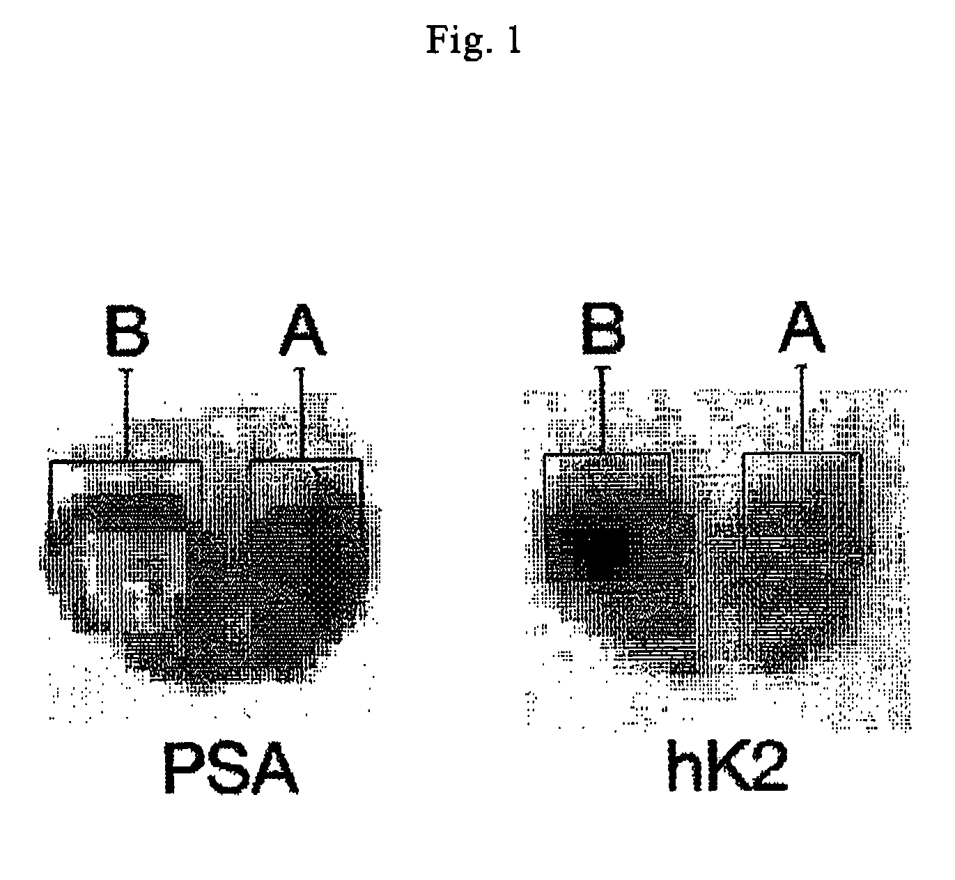
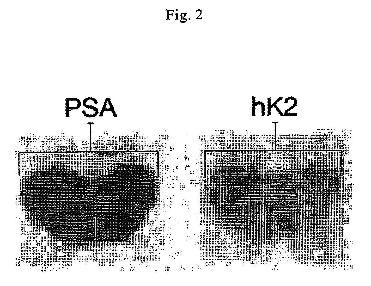
[0026]The following description focuses on embodiments of the present invention applicable to a diagnostic method of prostatic cancer. However, it will be appreciated that the invention is not limited to this application but may be applied to many other medical examinations, and diagnostic investigations, including for example lymph gland metastasis, post operative examinations, and examinations during or after radiation, cytostatic, and androgen treatments. In respect of diagnostic investigation of metastasis the metastases will be visible in lymph glands and lymph vessels, since PSA and hK2 pass these regions.

[0027]In an embodiment of the invention, antibodies that are specific for PSA and are labelled with a tracer are then injected into the body, such as intravenously. Then the tracer labelled antibodies, that are specific for PSA, bind to tissues that produce corresponding antigens, in this case PSA. The biologic structures, to which the tracer labelled PSA specific antibodies ...


PUM


Abstract

Methods for diagnosing prostate cancer, and differentiate prostate cancer from other prostate complications, and use of said method, and diagnosing and monitoring lymph gland metastasis, post operative examinations, and examinations during or after radiation, cytostatic, and androgen treatments are disclosed. The methods comprise injecting tracer-labelled PSA or hK2 specific antibodies, visualising PSA or hK2 producing tissue with the aid of a visualization method, and diagnosing prostate cancer from the difference in visualization.

Description

FIELD OF THE INVENTION[0001]This invention pertains in general to the field of a diagnostic method, which method diagnoses and distinguishes prostate cancer from other prostate complications, such as benign prostatic hyperplasia, and prostatitis. More particularly the invention relates to the use of said method.BACKGROUND OF THE INVENTION[0002]Prostate cancer is at the present time the most common form of cancer among men. The prostate is a walnut sized gland in men that produces fluid that is a component in semen. The prostate has two, or more, lobes, or sections, enclosed by an outer layer of tissue. The prostate is located in front of rectum and just below the urine bladder, and surrounds the urethra.[0003]The occurrence of prostate cancer is highest in the northwestern part of Europe and in the United States. The growth of the tumour is usually a procedure that takes place during a long period of time. Prostate cancer is normally a mild form of cancer. In fact, the majority of m...

Claims


Application Information

Patent Timeline
Patent Type & Authority Patents(United States)
IPC IPC(8): A61K49/00
CPCG01N2333/96455G01N33/57434C07K16/3069A61P35/00
Inventor ULMERT, DAVID
Owner JANSSEN BIOTECH INC
Learn More