System and method for acquiring magnetic resonance imaging (MRI) data

a magnetic resonance imaging and data acquisition technology, applied in the field of magnetic resonance imaging (mri), can solve problems such as mismatch between the actual image acquisition region and the anatomy of the patien

Active Publication Date: 2014-06-03
KONINK PHILIPS ELECTRONICS NV
View PDF20 Cites 26 Cited by
  • Summary
  • Abstract
  • Description
  • Claims
  • Application Information

AI Technical Summary

Benefits of technology

[0021]In this way, the proposed method achieves inter-scan prospective motion control which enables widespread use of shared geometries owing to the inventive approach of correcting predefined geometries prior to performing subsequent scans with the same geometry.
[0037]According to a variant of the inventive method, the geometry transformation matrix is derived by registering data from a first scan relative to data from a second scan, as described in prior art document WO 2004 / 052206 A1 the respective disclosure of which is incorporated herein by reference. In a corresponding embodiment of the MRI system according to the present invention there is provided a registration unit adapted to register data from a first scan relative to data from a second scan, and the control system further comprises geometry transformation generating means for deriving from said registration a transformation for use by the geometry updating means in updating the at least one predefined scan geometry. Alternatively, the geometry transformation matrix can be derived by registering data from a scan relative to an anatomical reference, i.e. an anatomical landmark of the patient's body, which can be retrieved easily during comparison of scan images regardless of a variation of MR settings, e.g. contrast. In a corresponding embodiment of the MRI system according to the present invention there is provided a registration unit adapted to register data from a scan relative an anatomical reference, and the control system further comprises geometry transformation generating means for deriving from said registration a transformation for use by the geometry updating means in updating the at least one predefined scan geometry.
[0041]In still another embodiment of the method according to the invention it is proposed that the above-defined step c) is performed upon reconstruction of each scan in a set of scans. In this way the inventive method ensures continuous geometry updating which results in an optimum coverage of the intended ROI.

Problems solved by technology

If the patient moves between scans there would be a mismatch of the actual image acquisition region and the patient's anatomy.

Method used


Image

  • System and method for acquiring magnetic resonance imaging (MRI) data
  • System and method for acquiring magnetic resonance imaging (MRI) data
  • System and method for acquiring magnetic resonance imaging (MRI) data

Examples


Embodiment Construction

[0047]The following detailed description of the invention refers to the accompanying drawings. The same reference numerals may be used in different drawings to identify the same or similar elements.

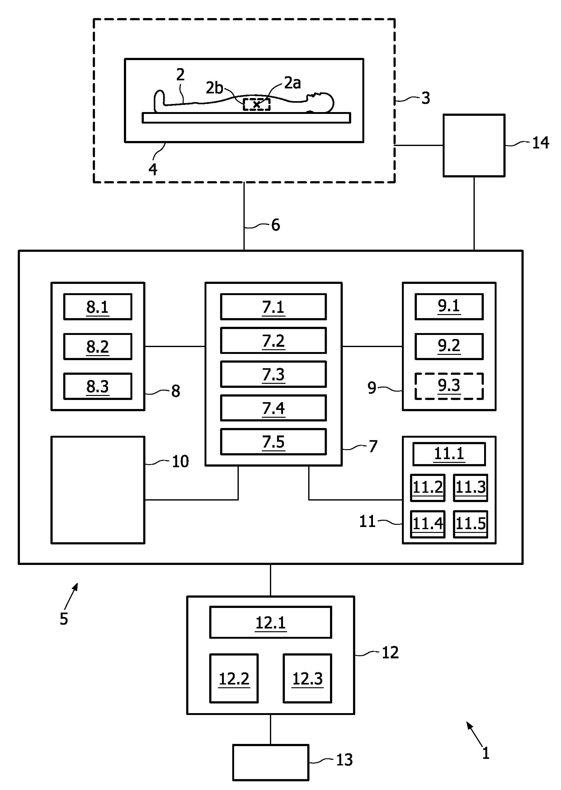
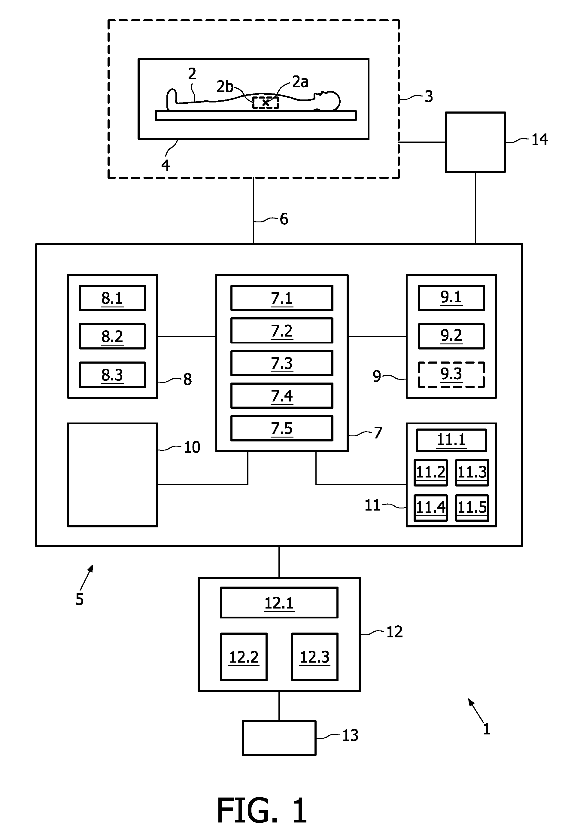
[0048]FIG. 1 shows a block diagram of an MRI system 1 for acquiring image data from an examination subject 2, e.g. a patient. The system 1 comprises an MRI examination chamber or magnet room 3, inside which the patient is positioned for examination. Within the examination chamber 3, the system 1 comprises a magnet / coil device 4 arranged around the patient 2. The magnet / coil device 4 includes various functional units (not shown) adapted for subjecting the patient 2 to a specific magic field used to elicit NMR signals from a particular location 2a (denoted “X”) inside the patient 2. Said functional units are generally known to a person skilled in the art and regularly comprise polarising magnets, shim coils, RF coils, and gradient coils. Said particular location 2a is usually extended by me...


PUM


Abstract

A method for acquiring image data from a patient with a magnetic resonance imaging (MRI) system. The proposed method comprises the steps of: a) predefining a number of scan geometries for acquiring the image data from at least one region of interest (ROI) relative to the patient, b) performing at least one scan for acquiring the image data in accordance with at least one of the predefined scan geometries, c) analysing in the image data a position of the region of interest to detect a deviation from the at least one predefined scan geometry, d) changing the at least one predefined scan geometry if said deviation exceeds a predetermined threshold value, and e) repeating steps b) to d) until a predetermined number of scans has been performed. Thus, by means of the proposed method the utility of such predefined scan geometries is greatly enhanced.

Description

TECHNICAL FIELD OF THE INVENTION[0001]The present invention relates generally to Magnetic Resonance Imaging (MRI). More specifically, the present invention relates to a method and a system for acquiring MR image data from a patient using predefined scan geometries.BACKGROUND OF THE INVENTION[0002]Utilising nuclear magnetic resonance (NMR) has become a standard procedure for noninvasively acquiring image data from inside a patient's body, e.g. the brain, by means of clinical MRI equipment. In this context, a technique using magnetic field gradients is employed which permits to obtain NMR signals (constituting NMR data) from specific locations in the patient. Typically, the region which is to be imaged (region of interest, ROI) is scanned in planes by a sequence of measurement cycles. The resulting NMR data are digitized and further processed to reconstruct the image using one of a variety of reconstruction techniques known to a person skilled in the art.[0003]Prior art methods and sy...

Claims


Application Information

Patent Timeline
Patent Type & Authority Patents(United States)
IPC IPC(8): A61B5/055
CPCG01R33/56509
Inventor VAN, DEN, BRINK, JOHAN, SAMUEL
Owner KONINK PHILIPS ELECTRONICS NV
Why Eureka
Try Eureka