Near infrared fluorogen and fluorescent activating proteins for in vivo imaging and live-cell biosensing

a technology of in vivo imaging and live cells, applied in the field of fluorogens and biosensors, can solve the problems of partially restricting the application of fap technology, and achieve the effects of reducing the excitation and detection of traditional fluorescent probes

Active Publication Date: 2015-05-05
CARNEGIE MELLON UNIV
View PDF12 Cites 0 Cited by
  • Summary
  • Abstract
  • Description
  • Claims
  • Application Information

AI Technical Summary

Benefits of technology

[0012]A novel fluorescent tag was developed, based upon a genetically targeted fluorogen-activating protein and cognate fluorogenic dye that yields emission with a peak at 733 nm exclusively when complexed as a “fluoromodule”. This tool improves substantially over previously described far-red / NIR fluorescent proteins in terms of brightness, wavelength, and flexibility by leveraging the flexibility of synthetic chemistry to produce novel chromophores. Indeed, the basic aromatic fluorogen structure can be modified with, for example, an alkyl group, and / or with charged groups on a linker. These modifications can be used to control water solubility and the ability of the fluorogen to cross, or be blocked by, biological membranes. This technology was used for in vivo imaging applications which resulted in robust, in vivo detection of FAPs binding the novel fluorogen SKC602 in living mice. These results suggest the new fluoromodule can bypass previous limitations imposed for fluorescence-based studies within tissues, intact organs, and living organisms.
[0013]The development of these new fluorogen structures and cognate FAPs that absorb and emit at wavelengths longer that the previous fluorogen / FAPs described above will allow for a number of new applications and improvements upon existing ones. First, fluorescence detection in the Near-Infrared region (NIR, >700 nm-2500 nm) permits detection of fluorescent structures deeper in tissues. This is because hemoglobin and other naturally occurring biomolecules absorb light strongly in the visible light region, limiting the excitation and detection of traditional fluorescent probes deep in tissues. However, longer NIR wavelengths penetrate tissues more deeply, allowing more efficient excitation of the probe, as well as more effective capture of the emitted light.
[0014]NIR detection assays that can be executed at longer wavelengths will result in less false-positive or false-negative events caused by fluorescent drug candidates. Additionally, there is less biological background fluorescence at longer wavelengths which would subsequently increase detection sensitivity.
[0015]Additionally, a new detection range can be used in combination with FAP-Fluorogens that are detected at multiple wavelengths in the visible range of the spectrum allowing the simultaneous detection of several biological properties of individual living or fixed cells. Multi-parameter detection not only speeds the amount of data per unit time but allows correlation of multiple parameters. This has great importance in studies of correlated physiological and biochemical activities in cells.

Problems solved by technology

This partially limits the application of the FAP technology, particularly towards the interrogation of tissue slices, organs, and whole animals, which absorb and scatter visible light.

Method used


Image

  • Near infrared fluorogen and fluorescent activating proteins for in vivo imaging and live-cell biosensing
  • Near infrared fluorogen and fluorescent activating proteins for in vivo imaging and live-cell biosensing
  • Near infrared fluorogen and fluorescent activating proteins for in vivo imaging and live-cell biosensing

Examples


example 1

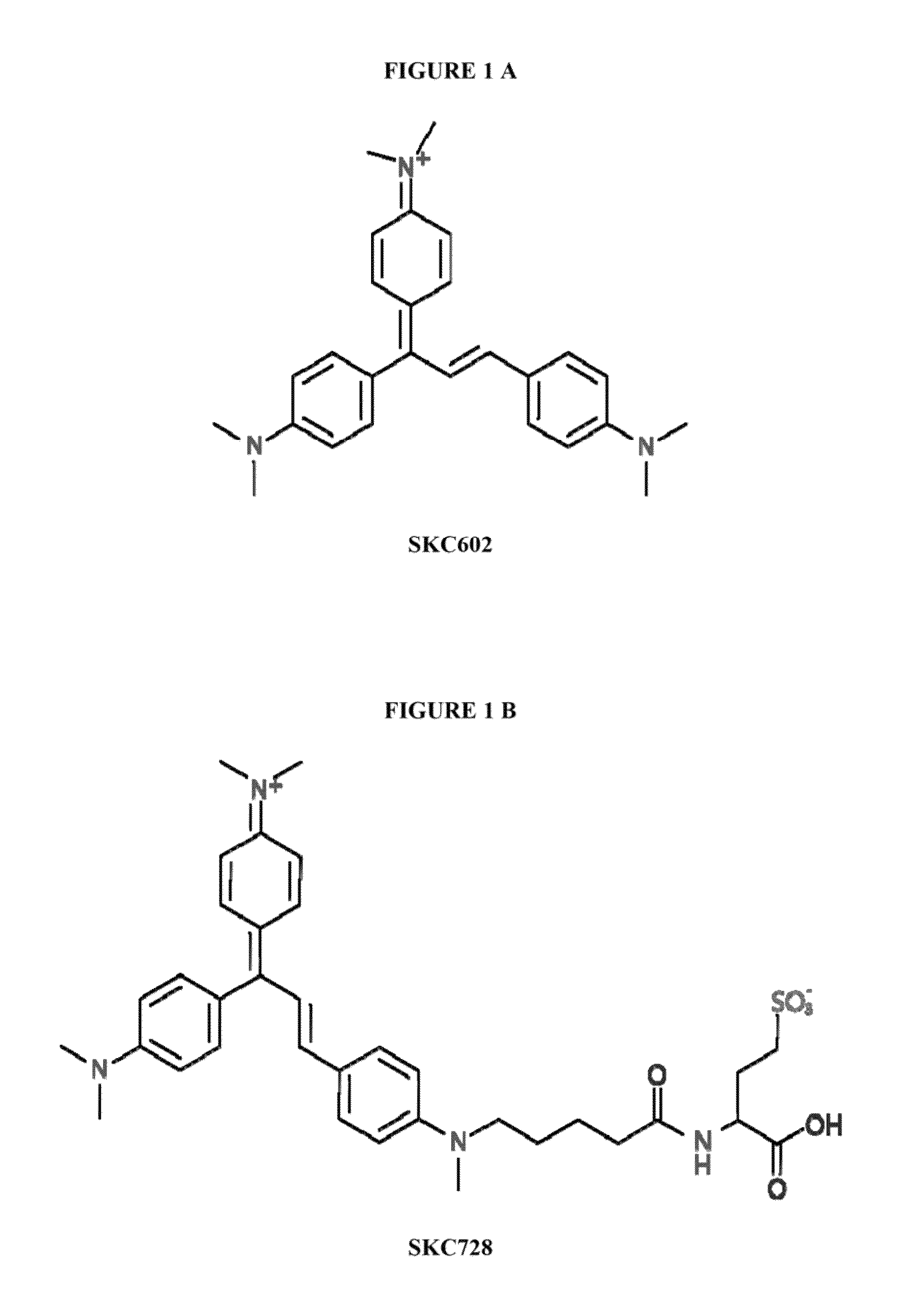
Synthesis of [(E)-N-(4-(1,3-bis(4-(dimethylamino)phenyl) allylidene)cyclohexa-2,5-dien-1-ylidene)-N-methylmethanaminium] (SKC602)

[0056]

[0057]4-Dimethyl amino benzaldehyde (149 mg, 1 mmol) and 4,4′-vinylidene bis(N,N-dimethylaniline) (300 mg, 1.12 mmol) were dissolved in 5 ml acetic anhydride. 200 μl of 60% perchloric acid was added slowly. The mixture was refluxed for 3 hour under stirring and cooled to room temperature. The mixture was precipitated by diethyl ether, and the blue residue was purified by silica gel column chromatography using 10% methanol in chloroform to give a 242 mg deep blue solid as the product. MW C27H32N3+398.56 g / mol (61% yields).

[0058]1H-NMR (CD3OD): δ 7.72-7.85 (m, 2H); 7.53 (d, 2H); 7.45 (d, 1H); 7.12 (d, 1H); 6.52-6.98 (m, 8H); 3.24 (d, 12H); 2.9 (m, 6H). ESI-MS (+): 398.3.

example 2

Synthesis of SKC638

[0059]

[0060]4-(dimethylamino)cinnamaldehyde (355 mg, 2 mmol) and 4,4′-vinylidene bis(N,N-dimethylaniline) (585.37 mg, 2.2 mmol) were dissolved in 10 ml acetic anhydride and 300 μl of HClO4 was added. The mixture was refluxed for 3 hour under stirring and cooled to room temperature. The mixture was precipitated by diethyl ether, and the residue was purified by silica gel column chromatography using 5% methanol in chloroform to give a 202 mg solid (24% yields) as the product. MW: C29H34N3+424.27 g / mol; ESI-MS (+): 424.67.

example 3

Synthesis of [Ethyl 5-(methyl(phenyl)amino)pentanoate] (SKC659)

[0061]

[0062]5.52 ml (48 mmol) of N-Methyl aniline, 8 ml Bromovalerate (50.2 mmol) and 2,6-lutidine (5.82 ml, 50.2 mmol) were taken in 100 ml acetonitrile and the reaction was refluxed for 24 hour under stirring. Acetonitrile was removed under vacuum and residues were dissolved in ether. The organic phase was washed with water. After drying over magnesium sulfate and filtering, the solvent was removed to give a brown liquid. The residue was purified by flash chromatography on silica gel using a 10% ethyl acetate in hexane as eluent gave 10.1 g light yellow oil as product. MW C14H21NO2 235.32 g / mol (yields 90%).

[0063]1H-NMR (CDCl3): δ 7.25 (m, 2H); 6.72 (m, 3H); 4.15 (m, 2H); 3.35 (m, 2H); 2.94 (s, 3H); 2.35 (m, 2H); 1.67 (m, 4H); 1.27 (m, 3H). ESI-MS (+): 235.62


PUM

PropertyMeasurementUnit
wavelengthsaaaaaaaaaa
temperatureaaaaaaaaaa

Abstract

Tissue slices and whole organisms offer substantial challenges to fluorescence imaging. Autofluorescence and absorption via intrinsic chromophores, such as flavins, melanin, and hemoglobins, confound and degrade output from all fluorescent tags. An “optical window,” farther red than most autofluorescence sources and in a region of low hemoglobin and water absorbance, lies between 650 and 900 nm. This valley of relative optical clarity is an attractive target for fluorescence-based studies within tissues, intact organs, and living organisms. Novel fluorescent tags were developed herein, based upon a genetically targeted fluorogen activating protein and cognate fluorogenic dye that yields emission with a peak at 733 nm exclusively when complexed as a “fluoromodule”. This tool improves substantially over previously described far-red / NIR fluorescent proteins in terms of brightness, wavelength, and flexibility by leveraging the flexibility of synthetic chemistry to produce novel chromophores.

Description

PRIORITY[0001]This application claims the benefit of U.S. Provisional Patent Application Ser. No. 61 / 850,872 filed Feb. 25, 2013 and Ser. No. 61 / 851,789 filed Mar. 13, 2013; the contents of each of which are incorporated by reference herein in their entirety.GOVERNMENT RIGHTS[0002]This invention was made with government support under the National Institute of Health 5U54RR022241. The government has certain rights in this invention.SEQUENCE LISTING[0003]This application includes a Sequence Listing submitted via EFS-Web in computer readable form contained in a 82 Kb byte file entitled 130104_ST25(New).txt created on Jun. 18, 2013 and amended on Oct. 29, 2014 using an IBM-PC machine format, which is hereby incorporated herein by reference in its entirety.TECHNICAL FIELD[0004]The present disclosure is directed to fluorogens and biosensors comprising fluorogens and fluorogenic activating peptides, methods of producing the fluorogens and biosensors, and methods of using the same.BACKGROUN...

Claims


Application Information

Patent Timeline
Patent Type & Authority Patents(United States)
IPC IPC(8): C07K16/44A61K49/00C07C251/30C07C309/18C07D207/46G01N21/64
CPCG01N21/6486A61K49/0021C07D207/46C07C309/18C07C251/30C07K16/44C07K2317/515A61K49/0023C09B23/0008C09B23/04C09B23/06C09B23/08
Inventor CHAKRABORTY, SUBHASISH K.ZHANG, MINGRUIWAGGONER, ALAN S.
Owner CARNEGIE MELLON UNIV
Why Eureka
Try Eureka