Methods for detecting antibodies
a detection method and antibody technology, applied in the field of methods for detecting antibodies, can solve the problems of affecting the ability of patients, unable to obtain reliable detection of ada in samples, etc., and achieve the effect of improving the patient's response or the efficacy of treatmen
- Summary
- Abstract
- Description
- Claims
- Application Information
AI Technical Summary
Benefits of technology
Problems solved by technology
Method used
Image

Examples
examples
[0124]In the experimental section, every dosage is quantitative, calibrated with regard to standards. Dosage of TNF (pg / ml) is made with the help of the international standard. Dosage of the drug (μg / ml) is made thanks to the concentration value given by the pharma company. Dosage of ADA (ng / ml) is made using a rabbit polyclonal antibody as a standard. Rabbits (origin Hyla) are immunized with Fab or Fab′2 fragments of the anti-TNF immunoglobulins or with human P75 TNF receptor. Quantification of the ADA human standard is obtained by the method of the biuret (dosage of protein by colorimetry).
example a
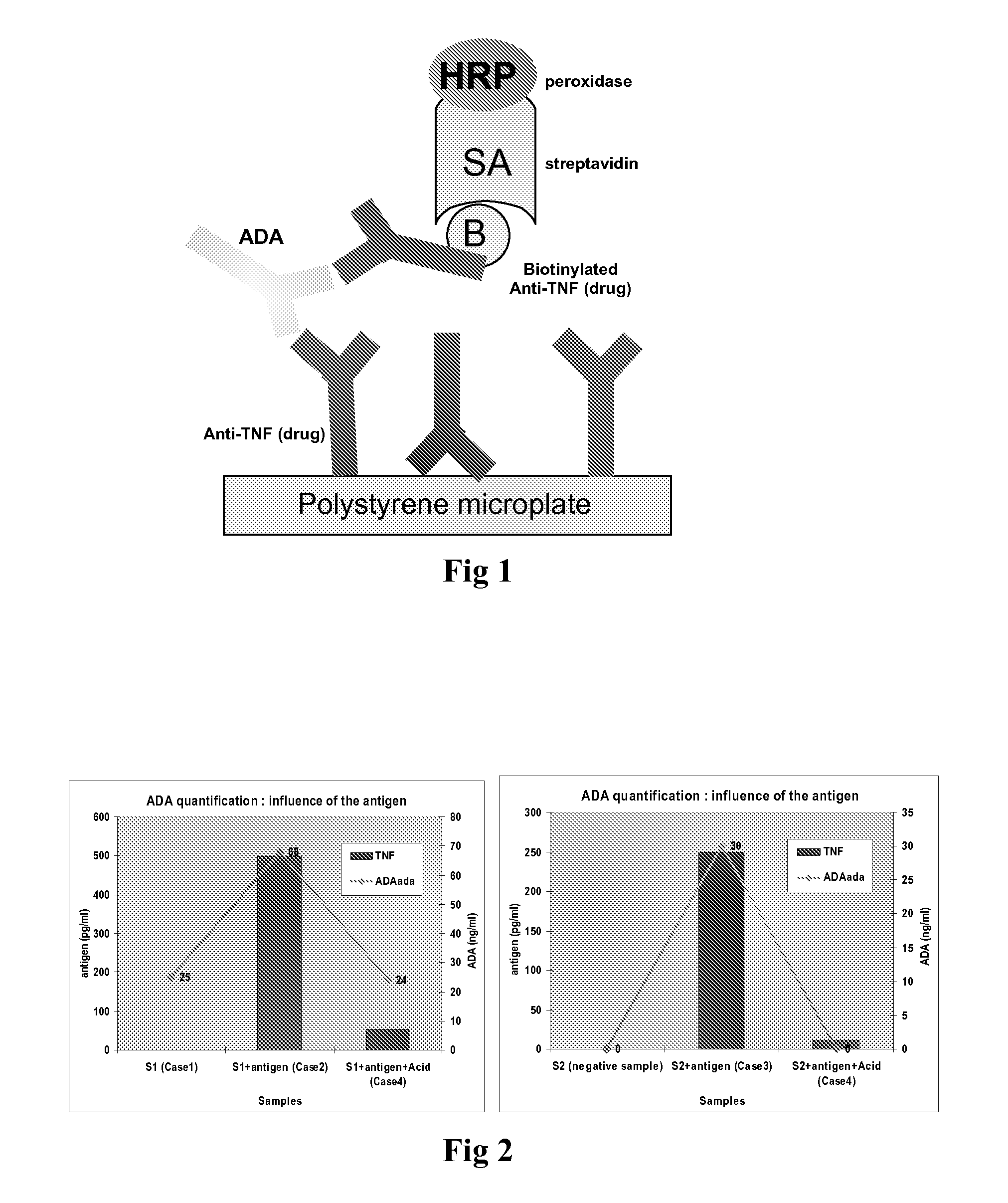
Method of Detecting ADA
[0125]The drug is coated onto a polystyrene microtiter plate.[0126]First, an acid solution, such acid acetic solution, is added to the sample. The acidified sample is incubated at room temperature for approximately 5 minutes.[0127]Then, a basic solution, such as TRIS solution, is added to the acidified sample to neutralize acid solution.[0128]Then, sample (diluted or not) is added to the antibody coated well, which allows to bind. After incubation, unbound proteins are removed by washing.[0129]Biotinylated drug is added. After incubation, unbound antibodies are removed by washing.[0130]Then horseradish peroxidase labelled streptavidin is added. The streptavidin binds to the complex formed with biotinylated drug. After incubation, the wells are washed again to eliminate any excess of conjugate.[0131]The bound enzyme is revealed by addition of substrate TMB (3,3′,5,5′ tetramethylbenzidin). The colour intensity is proportional to the amount of anti-Etanercept ant...
example b
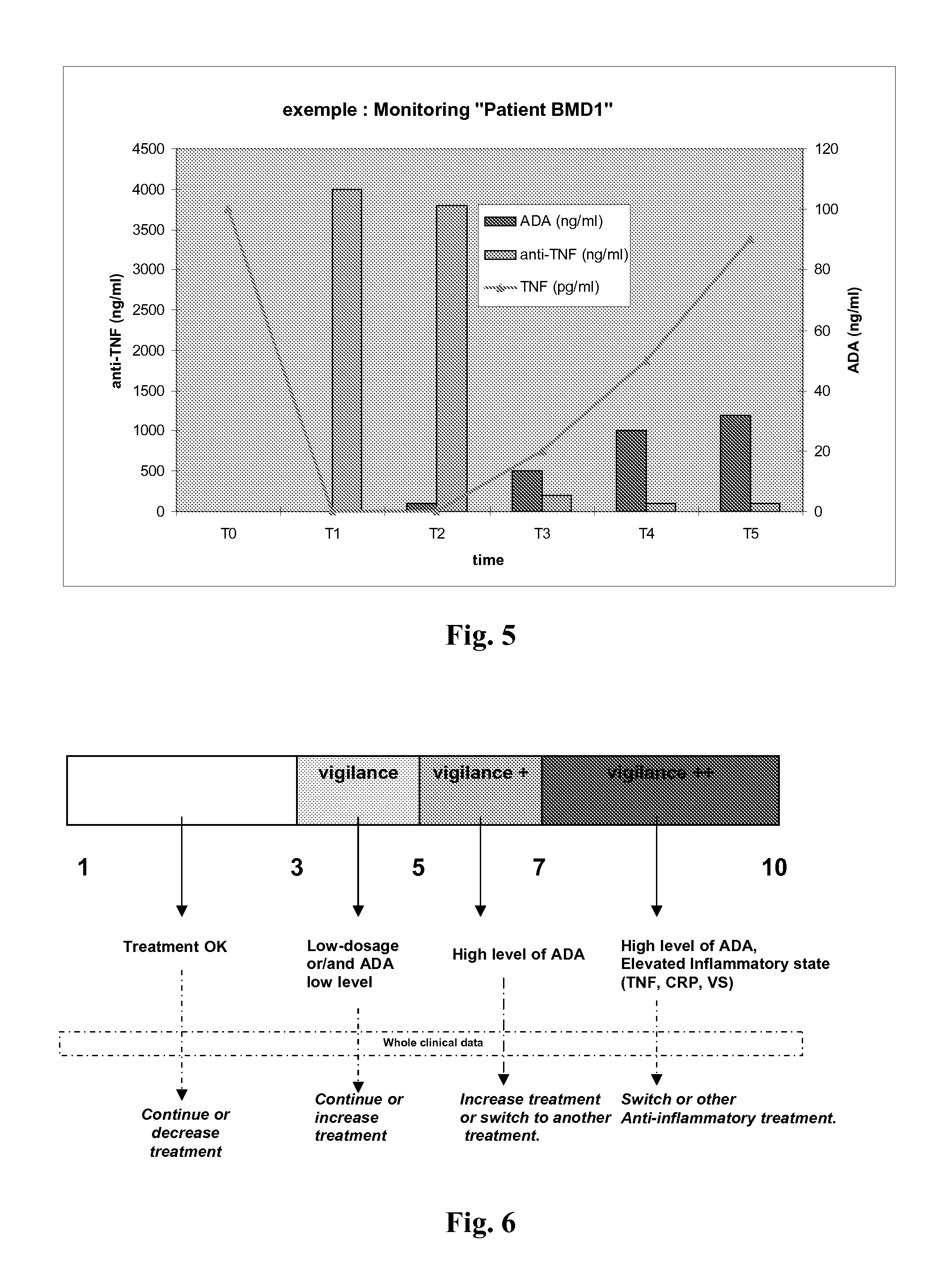
Method and Kit for Monitoring Patients Treated with Etanercept
[0137]The test should be performed on serum or on plasma. TNFα being unstable in solution, it is preferred to use samples immediately after blood taking. If determination is not performed immediately, samples should be frozen. To avoid any non-specific binding, samples which have been frozen for more than 6 months or which are cloudy, should be centrifuged and filtered.
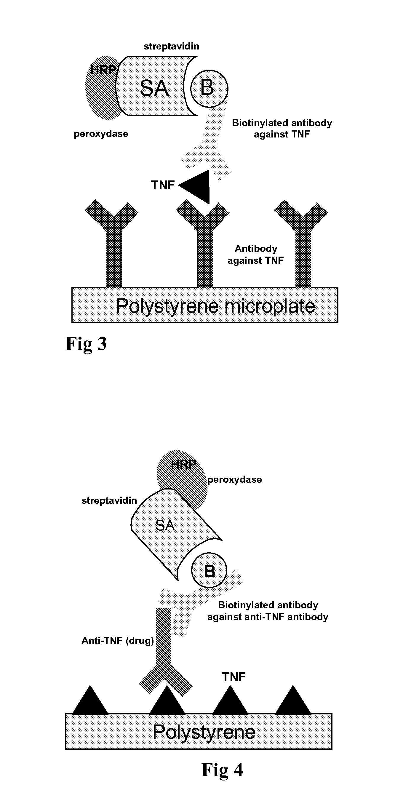
[0138]1. Dosage of TNFα
[0139]A monoclonal anti-TNFα antibody is coated onto a polystyrene microtiter plate (4 strips of 8 wells).[0140]First, the diluted sample is added to the antibody coated well, which allows to bind. After incubation, unbound proteins are removed by washing.[0141]Anti-TNFα biotinylated antibody is added. After incubation, unbound antibodies are removed by washing[0142]Then horseradish peroxydase labelled streptavidin is added. The streptavidin binds to the complex formed with biotinylated anti-TNFα antibodies. After incubation, the well...
PUM
| Property | Measurement | Unit |
|---|---|---|
| time | aaaaa | aaaaa |
| pH | aaaaa | aaaaa |
| affinity | aaaaa | aaaaa |
Abstract
Description
Claims
Application Information
 - R&D
- Intellectual Property
- Life Sciences
- Materials
- Tech Scout
- Unparalleled Data Quality
- Higher Quality Content
- 60% Fewer Hallucinations
Browse by: Latest US Patents, China's latest patents, Technical Efficacy Thesaurus, Application Domain, Technology Topic, Popular Technical Reports.
© 2025 PatSnap. All rights reserved.Legal|Privacy policy|Modern Slavery Act Transparency Statement|Sitemap|About US| Contact US: help@patsnap.com