
Nanoporous metal multiple electrode array and method of making same

a technology of nanoporous metal and multiple electrodes, applied in the field of multiple electrode arrays, can solve the problems of poor functional coating adhesion, difficult to obtain high spatial resolution, complicated fabrication techniques, etc., and achieve the effects of improving electrode-electrolyte impedance, low impedance, and high surface area-to-volume ratio

Inactive Publication Date: 2015-06-30
THE GENERAL HOSPITAL CORP
View PDF9 Cites 18 Cited by
  • Summary
  • Abstract
  • Description
  • Claims
  • Application Information

AI Technical Summary

Benefits of technology

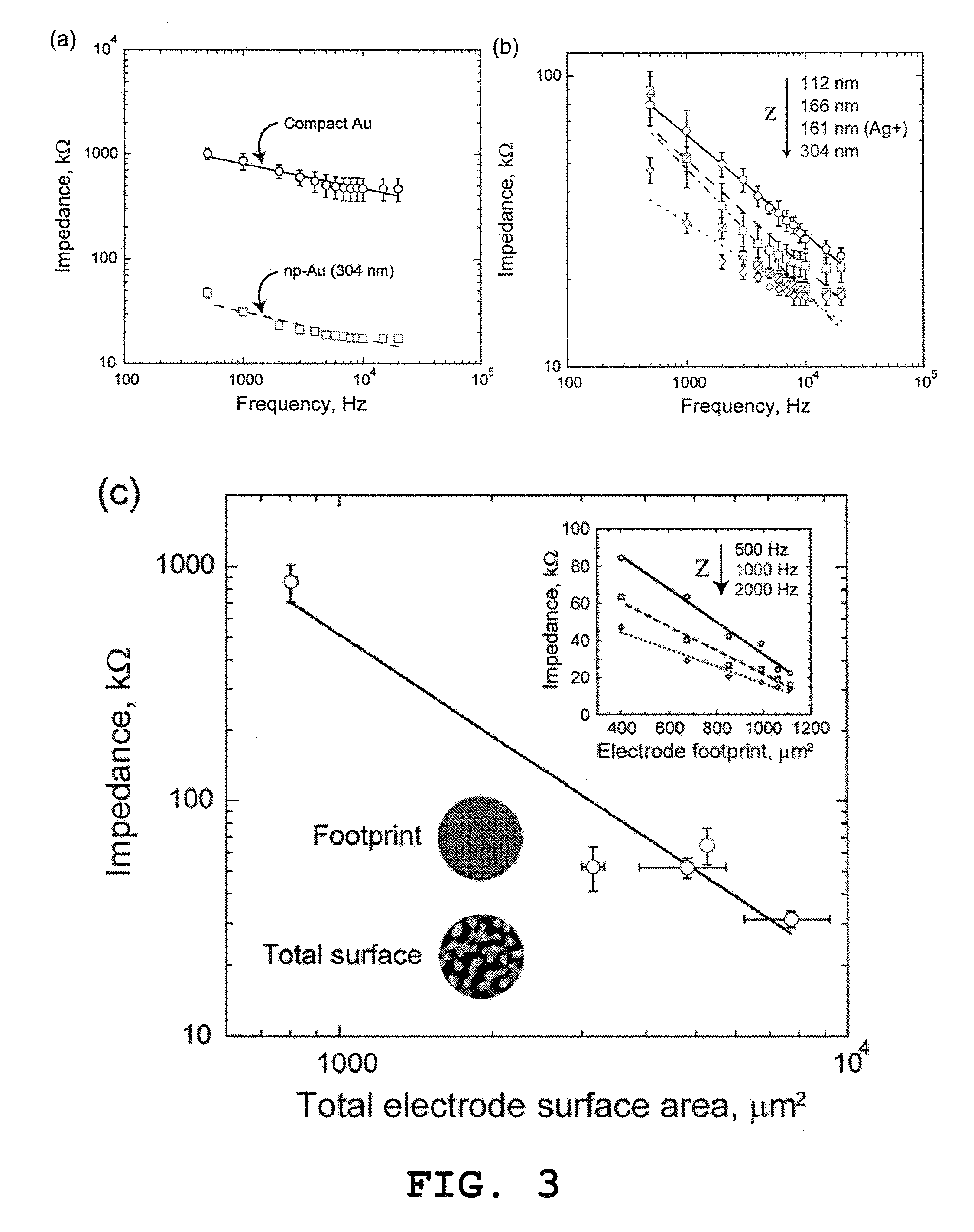
[0013]The disclosed nanoporous metal MEA and related method of making has many benefits. The method utilizes simple microfabrication techniques in a unique way to create a novel structure having much sought after, but previously unachieved, impedance and spatial resolution qualities. The resultant multiple electrode arrays exhibit lower impedance than the present state-of-the-art platinum black electrodes.
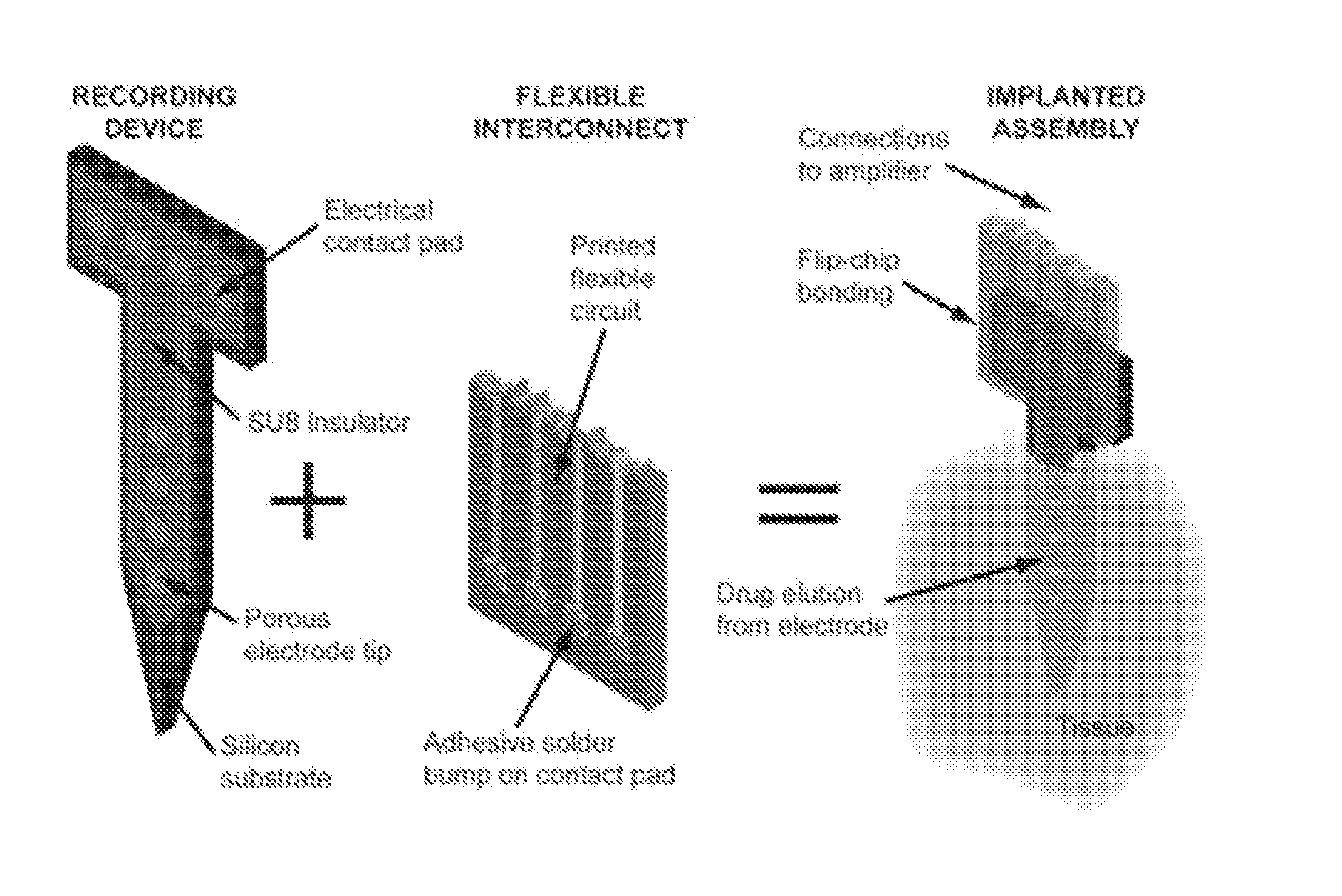
[0015]In one preferred form of the device, the electrode tips include a gold material so that a nanoporous gold multiple electrode array is formed (which may be referred to as a “np-Au MEA”). The np-Au MEA has a thiol-based gold surface chemistry that may further allow advanced functionalization of the electrodes (e.g., immobilization of extra-cellular matrix proteins). Moreover, the high surface area-to-volume ratio of self-assembled nanoporous gold can result in more than a 25-fold improvement in electrode-electrolyte impedance, where at 1 kHz, 850 kΩ impedance for conventional Au electrodes is reduced to 30 kΩ for np-Au electrodes. Low impedance provides superior signal-to-noise ratio for detection of neural activity in noisy environments. The fabricated np-Au MEAs may be used to measure electrophysiological activity in vitro or in vivo.

Problems solved by technology

Although low electrode impedance is a figure of merit for sensitive detection of neural electrical activity and numerous studies have aimed to reduce impedance, fabrication of a robust multiple electrode array that significantly reduces impedance while obtaining high spatial resolution has proven to be a challenge.
Unfortunately, to date, most efforts to do so have been tethered by a combination of poor functional coating adhesion, complicated fabrication techniques, and poor fabrication repeatability.

Method used


Image

  • Nanoporous metal multiple electrode array and method of making same
  • Nanoporous metal multiple electrode array and method of making same
  • Nanoporous metal multiple electrode array and method of making same

Examples


example i

[0049]Various materials used in preparation of the nanoporous gold multiple electrode arrays were obtained as follows. Glass slides 75 mm×50 mm were purchased from VWR (West Chester, Pa.). Gold, silver, and chrome targets were all 99.95% pure and obtained from Kurt J. Lesker (Clairton, Pa.). Ethanol, methanol, acetone, sulfuric acid, hydrogen peroxide, hexamethyldisilazane (photoresist adhesion promoter) were obtained from Sigma-Aldrich (St. Louis, Mo.). AZ400K developer and AZ4330 positive photoresist (PR) were obtained from Clariant Corporation (Somerville, N.J.). SU8-2 and Edge Bead Removal solution (SU8 developer) were obtained from Microchem Corporation (Newton, Mass.). Polydimethylsiloxane (PDMS) kit was obtained from Sylgard 184, Dow Corning (Wilmington, Mass.). Silicon wafers were obtained from Silicon Quest International, Inc. (Santa Clara, Calif.). Impedance measurements and field potential recordings were carried out in artificial cerebrospinal fluid (ACSF), composed of 1...

example ii

[0079]To help establish the efficacy of various drug or pharmaceutical coatings on a nanoporous metal material, we developed an immunostaining protocol to visualize astrocytes, microglia, and cell proliferation, which are key indicators of a foreign body response. By administering such a protocol to test tissue samples, the integrity of a tissue sample can be quantitatively assessed.

[0080]In order to test the effect of a classical anti-inflammatory drug, dexamethasone, in gliosis, we dissected 350 μm-thick hippocampus slices from postnatal day 7 Sprague-Dawley rat pups. Organotypic slices have been shown to provide a physiologically and pharmacologically relevant model, as they preserve the cytoarchitecture of the brain and provide a means to identify pharmaceuticals for in vivo validation. In addition, the tissue damage due to slice preparation partially mimics the damage induced by neural probe insertion.

[0081]The slices were seeded in a serum-containing medium (1:1:2 horse serum,...

example iii

[0084]Having established a protocol for the evaluation of glial scarring as described in Example II, in separate trials we used fluorophore-conjugated primary antibodies: anti-glial fibrillary acidic protein (anti-GFAP) for astrocytes (red) and anti-BrdU (infra-red), and subsequently imaged CA3 and DG regions of the slices using a fluorescent confocal microscope at 7× magnification and clearly identified BrdU and GFAP positive cells. For live microglia staining, we applied fluorophore-conjugated Isolectin-B4 with the regular medium for 1 hour and imaged slices with a confocal microscope. For image analysis we used Fiji (ImageJ) to determine the number of proliferating cells (BrdU positive) and astrocyte reactivity (GFAP intensity).

[0085]Using the organotypic slices and immunostaining approaches, the effect of different pharmaceutical medium supplements were evaluated at various concentrations: (i) immunosuppressant rapamycin (0.1 and 1 μM), a mTOR signaling inhibitor that results in...


PUM

PropertyMeasurementUnit
porosityaaaaaaaaaa
porosityaaaaaaaaaa
impedanceaaaaaaaaaa

Abstract

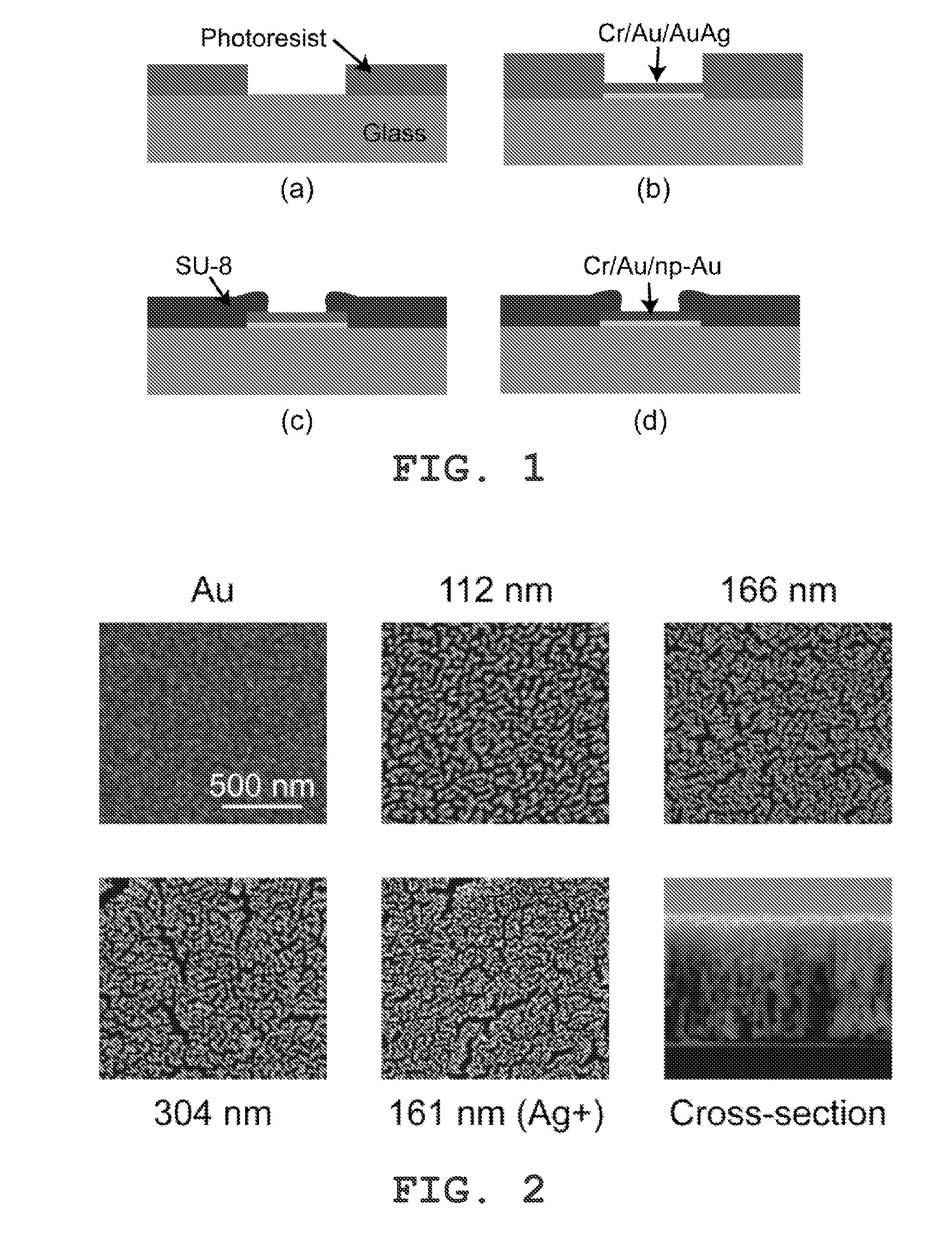
A method is disclosed for fabricating a low-impedance nanoporous metal multiple electrode array for measuring electrophysiology activity. A patterned photoresist is applied to a substrate, in which the patterned photoresist corresponds to a pattern of the nanoporous metal multiple electrode array. A metal alloy including a sacrificial alloying element is deposited in the pattern of the nanoporous metal electrode array. The patterned photoresist is removed to expose the metal alloy as deposited. At least part of the sacrificial alloying element is removed from the metal alloy to create nanoporous metal electrode tips thereby forming the nanoporous metal multiple electrode array. The resultant nanoporous metal multiple electrode array has improved impedance characteristics in comparison to conventional multiple electrode arrays.

Description

CROSS-REFERENCE TO RELATED APPLICATION[0001]This patent application represents the national stage entry of PCT international application no. PCT / US2011 / 051388 filed on Sep. 13, 2011 which claims priority to U.S. provisional patent application Ser. No. 61 / 382,610 entitled “Nanoporous Metal Multiple Electrode Array and Method of Making Same” filed on Sep. 14, 2010 and to U.S. provisional patent application Ser. No. 61 / 475,893 entitled “Neural Electrode Array and Methods of Using Same” filed on Apr. 15, 2011. The full contents of those applications are incorporated by reference as if set forth in their entirety herein.STATEMENT REGARDING FEDERALLY SPONSORED RESEARCH[0002]This invention was made with the government support under P41-EB002503 and F32-MH079662 awarded by the National Institutes of Health and DMI-0507023 awarded by the National Science Foundation. The government has certain rights in the invention.BACKGROUND OF THE INVENTION[0003]This invention relates to multiple electrod...

Claims


Application Information

Patent Timeline
Patent Type & Authority Patents(United States)
IPC IPC(8): A61B5/0492A61B5/04G01N33/483H01B7/30A61B5/296
CPCH01B7/30A61B5/0492A61B5/04001G01N33/4836A61B5/24A61B5/296
Inventor YARMUSH, MARTIN L.SEKER, ERKINBERDICHEVSKY, YEVGENY
Owner THE GENERAL HOSPITAL CORP
Learn More