
Occlusion device for vascular surgery

a vascular surgery and occlusion device technology, applied in the field of vascular surgery, can solve the problems of reducing the efficiency of compression, and preventing the entry site of a vascular prosthesis made of plastic (dacron, ptfe, etc.) from premature hardening of the haemostatic liquid

Active Publication Date: 2016-05-10
GEM SPA
View PDF100 Cites 0 Cited by
  • Summary
  • Abstract
  • Description
  • Claims
  • Application Information

AI Technical Summary

Benefits of technology

[0022]It is therefore an object of the present invention to provide an occlusion device for haemostasis of arterial entry sites and for endovascular treatment, which uses a quick setting haemostatic liquid, for example a surgical glue, preventing the haemostatic liquid from prematurely hardening in the device and causing it to block.

Problems solved by technology

In some cases, even by prolonging the compression the entry site cannot be closed, and a surgical treatment is necessary, which may involve further risks, discomfort, prolonged period in bed and resources waste.
Furthermore, in case of obese patients, the adipose layer that is located between the skin and the vascular entry site reduces the efficiency of the compression.
Moreover, an entry site on a vascular prosthesis that is made of plastic (Dacron, PTFE, etc.) cannot heal by the natural repair processes of an artery, and, on the contrary, it can only be closed by formation of a true plug made of blood that, after flowing out of the opening, coagulates through the tissues that surround the prosthesis, under the assistance of the compression force.
Such systems reduce the average treatment time.
However, the introduction of objects into the blood vessels may cause thrombosis related ischemia, embolism, arterial wall laceration, sensitization, allergic reaction.
Furthermore, the mechanical closure devices require a highly skilled operator, and / or the work of further assistant operators.
Therefore, these techniques do not provide a valid alternative to the traditional techniques.
Systems are also known for closing the entry site of an introducer sheath with exclusive use of a haemostatic liquid, in particular collagen, to form clots that are involved in a natural coagulation mechanisms; however, the haemostasis process is slow, and complications may arise such as blood suffusion, bleeding, pseudoaneurysms.
The methods that use quick setting surgical glues provide a dilution of the glue, but this prolongs the hardening time;with well known techniques, and also under echographic control, it is difficult to release such pure quick setting haemostatic liquid proximate to a vascular entry site without the risk of injecting it into the blood vessel, with such complications as thrombosis or embolisms.
The echographic control may not be always available when this kind of intervention is performed, because it requires experienced operators and in any case it requires more than one operator for closing the entry site.
This causes an unsteady positioning of the introducer sheath, which is particularly critical in the case of obese patients, where the distance between the skin and the blood vessel is higher than in normal patients.
Furthermore, the large transversal size of the device hinders the movement through the tissues, and “digs” an enlarged space about the introducer sheath in which both the released haemostatic liquid and the blood can accumulate without contributing to the entry site occlusion.
In any case, the device is not adapted to treat a quick-setting liquid, because the delivery mouth may be easily clogged.
In particular the device of US2008 / 45700 is adapted to treat bleeding diseases, for example, in the stomach and in the intestine, but it cannot be used neither for an endovascular intervention, nor for closing an arterial entry site.
Due to its high reactivity, a cyanoacrylate material would immediately polymerize when brought into contact with the blood, which would reduce the passage through the catheter, or even clog it, in such a way that the release of a significant amount of surgical glue would be impossible.
This is a rough method, by which the risk may arise of injecting the procoagulating substance into the vessel; for the same reason, the method and the apparatus cannot be used to safely release surgical glues, which are not cited in the patent application.
Furthermore, the document does not provide precise indications about the way the liquid is supplied at the entry site.
In any case, the device cannot be used to release a quick setting surgical glue proximate to an entry site, because the glue would come into contact too early with the nearby biological fluids which would block the device; furthermore, it does not allow an operator to determine the point of delivery, unless an echographic control is provided.
Similarly to any well-known syringe, a similar device does not allow providing a quick setting. in particular of a haemostatic liquid in a pure state, at an entry site or in an is operation region of an endovascular surgery.
Concerning the endovascular surgery, such as an embolization of arterial or venous districts, or the treatment of arterial walls dissections, the problem often arises of covering a long distance between the entry site, which is normally made in the artery of the arm or in the femoral artery, and the operation region.
When the use is desirable of a pure quick setting surgical glue, which would have a maximum efficiency and application speed, the problem arises of conveying the glue through a long duct, and of preventing at the same time any risks of contact between the glue and the blood until the remote vascular district is reached where the intervention must be made.

Method used


Image

  • Occlusion device for vascular surgery
  • Occlusion device for vascular surgery
  • Occlusion device for vascular surgery

Examples


Embodiment Construction

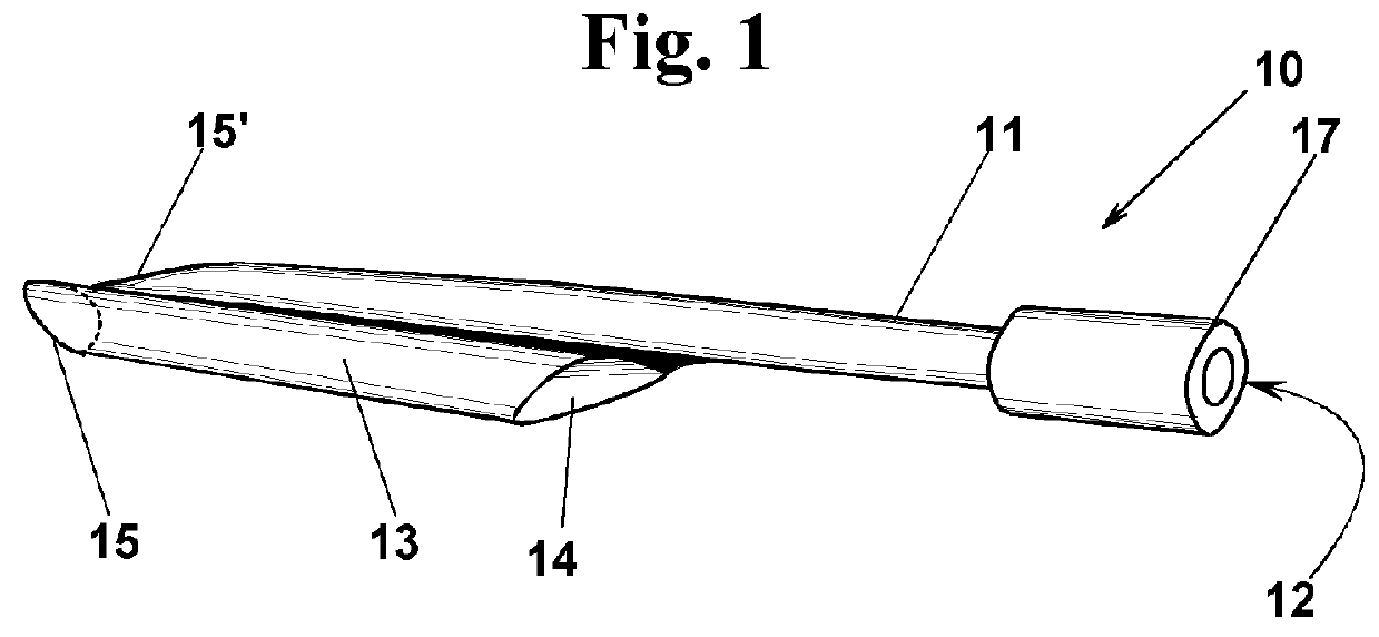
[0145]With reference to FIGS. 1, 2 and 3, a device 10 is described according to a first exemplary embodiment of the invention, for closing an entry site 2 in a blood vessel 1, or in a vascular prosthesis 1 (FIG. 3). A substantially cylindrical catheter introducer sheath 3, engages entry site 2. Device 10 (FIG. 1) comprises a duct 11 that has at one end an inlet port 12 that is associated with a luer-lock connection 17, or other connection suitable for connecting a source, not shown, of a surgical glue 9, for example a receptacle of a syringe. In the description, reference is made to a surgical glue, still remaining that the device can be advantageously used with any quick setting haemostatic liquid. At the opposite end, instead, an outlet mouth 18 (FIG. 2) is provided for surgical glue 9. For carrying out the haemostasis, outlet mouth 18 must be located at an operation region 4 (FIG. 3). A short tube 13, with a substantially cylindrical or slightly conical shape, and shorter than du...


PUM


Abstract

An occlusion device for vascular surgery, suitable for clogging treatments of vascular entry sites and for endovascular interventions such as embolizations of blood vessels, treatment of arteriovenous malformations or small aneurysms, arterial dissections and the like, by releasing in an operation region a quick setting surgical glue or haemostatic fluid, through an outlet mouth of a duct. The device prevents the surgical glue from contacting within the duct a patient's biological fluids, in particular blood, which would close the duct. In the case of clogging treatments of vascular entry sites, a backflow preventing device may be provided, preferably provided by a coupling device between the duct and an introducer sheath by which an outlet mouth is kept in contact to keep in a one-way fluid tight contact against the outer surface of the introducer sheath until an injection pressure P2 is applied to cause release of the surgical glue.

Description

FIELD OF THE INVENTION[0001]The present invention relates to a device for percutaneously closing apertures in walls of blood vessels and vascular prosthesis. Such apertures are made to perform diagnostic examinations, or to position surgical instruments for endovascular interventions. The invention relates also to a device for treating blood vessels haemorrhagic, dilatative or dissecting pathologies or arteriovenous malformations.[0002]In particular, the device can be used at the end of such a diagnostic procedure as arteriography or coronarography, or of such an endovascular intervention as angioplasty, stenting or embolization, which require a percutaneous puncture of an artery for positioning an introducer sheath in the vascular bed. In specific exemplary embodiments, the device can be used for treating pseudoaneurysms, for arterials or venous embolization, or for treating dissections of arterial walls.BACKGROUND OF THE INVENTION[0003]Many diagnostic and therapeutic procedures pr...

Claims


Application Information

Patent Timeline
Patent Type & Authority Patents(United States)
IPC IPC(8): A61B17/03A61M37/00A61B17/00A61B6/00
CPCA61B17/0057A61B17/00491A61B2017/0065A61B2017/00292A61B2017/00575A61B2017/00601
Inventor DEL CORSO, ANDREA
Owner GEM SPA
Learn More