Ultrasound endoscope

a technology of ultrasound endoscope and endoscope, which is applied in the field of ultrasound endoscope, can solve the problems of inability to pull out needles, inability to increase the raising angle of the treatment instrument of ultrasound endoscope, and inability to switch the ultrasound endoscope over to the ercp endoscop

Active Publication Date: 2016-07-05
OLYMPUS CORP
View PDF16 Cites 4 Cited by
  • Summary
  • Abstract
  • Description
  • Claims
  • Application Information

AI Technical Summary

Benefits of technology

[0011]An ultrasound endoscope according to an aspect of the present invention includes: an insertion portion configured to extend in a longitudinal direction and be inserted into a living body; an ultrasound transducer provided at a distal end portion of the insertion portion and emitting ultrasound sideward at a predetermined angle with respect to an insertion axis of the insertion portion; a first opening portion provided at the distal end portion of the insertion portion; a second opening portion provided at the distal end portion of the insertion portion at a position upper side than the first opening portion; a raising stand provided at the distal end portion of the insertion portion and movable between a first position proximate to the first opening portion and the second opening portion and a second position away from the first opening portion and the second opening portion; a first contact portion provided along the raising stand from a proximal end portion to a distal end portion and that comes into contact with a side face of a first treatment instrument protruding from the first opening portion to cause the first treatment instrument to extend in a first direction; and a second contact portion provided on the distal end portion of the raising stand and that comes into contact with a side face of a second treatment instrument protruding from the second opening portion to cause the second treatment instrument to extend in a second direction different from the first direction.

Problems solved by technology

During operation, the operation of switching the ultrasound endoscope over to the ERCP endoscope is complicated and also time-consuming.
However, since a puncture needle used during EUS-FNA is rigid, if the operator considerably bends the puncture needle beyond a certain limit, the needle itself may be broken or a bending tendency may be formed on the needle making it impossible to pull out the needle, and it is thereby not possible to increase the raising angle of the treatment instrument of the ultrasound endoscope.

Method used


Image

  • Ultrasound endoscope
  • Ultrasound endoscope
  • Ultrasound endoscope

Examples


first embodiment

(First Embodiment)

(Configuration of Ultrasound Endoscope System)

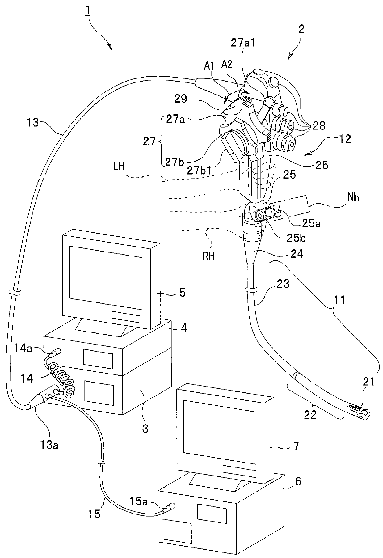
[0033]FIG. 1 is a configuration diagram illustrating an overall ultrasound endoscope system of the present embodiment.

[0034]An ultrasound endoscope system 1 is configured by including an ultrasound endoscope (hereinafter simply referred to as “endoscope”) 2, a light source apparatus 3, a video processor 4, a monitor 5 for displaying an optical image, an ultrasound observation apparatus 6 and a monitor 7 for displaying an ultrasound image.

[0035]The endoscope 2 includes an insertion portion 11, an operation portion 12 from which this insertion portion 11 extends and a universal cord 13 that extends from the operation portion 12. The insertion portion 11 extends in a longitudinal direction and configured to be inserted into a living body. The universal cord 13 is connected to the light source apparatus 3 via a scope connector 13a provided at a proximal end portion. A coiled scope cable 14 and an ultrasound signal cable 15 ...

second embodiment

(Second Embodiment)

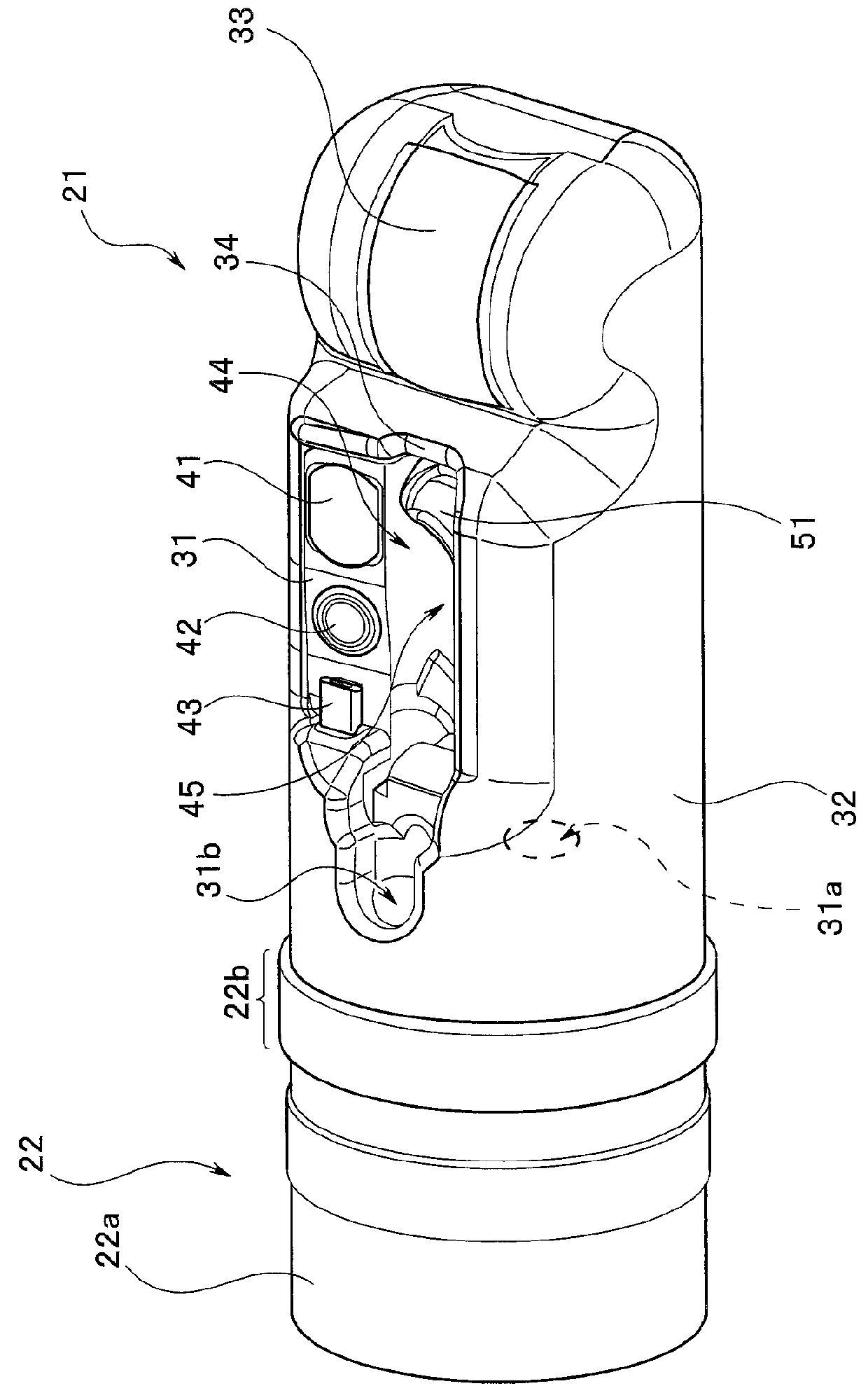
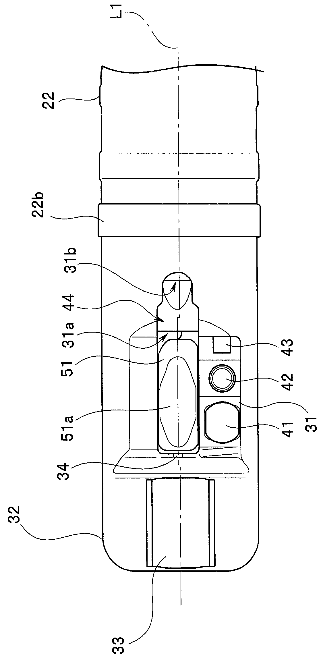
[0103]In the first embodiment, when the distal end portion 21 is seen from the distal end side, the two opening portions of the two channels in the distal end portion 21 are arranged along a direction from the bottom surface of the concave portion 45 toward the opening 44, that is, arranged side by side along the vertical direction. In the second embodiment, however, when the distal end portion 21 is seen from the distal end side, the two opening portions of the two channels in the distal end portion 21 are provided along a diagonal direction at a predetermined angle with respect to a direction from the bottom surface of the concave portion 45 toward the opening 44. That is, according to the second embodiment, in a plan view of the opening portion of the distal end portion of the insertion portion, the two opening portions of the two channels are arranged shifted in a direction orthogonal to the insertion axis of the distal end portion.

[0104]Note that in the prese...


PUM


Abstract

An ultrasound endoscope includes an insertion portion, an ultrasound transducer provided at a distal end portion of the insertion portion, a first opening portion provided at a distal end portion of the insertion portion, a second opening portion and a raising stand provided at the distal end portion of the insertion portion. Furthermore, the raising stand includes a first contact portion that comes into contact with a treatment instrument protruding from the first opening portion for causing the treatment instrument to extend in a first direction and a second contact portion that comes into contact with a treatment instrument protruding from the second opening portion for causing the treatment instrument to extend in a second direction different from the first direction.

Description

CROSS REFERENCE TO RELATED APPLICATION[0001]This application is a continuation application of PCT / JP2013 / 073986 filed on Sep. 5, 2013 and claims benefit of U.S. Provisional Patent Application No. 61 / 696,920 filed in the U.S.A. on Sep. 5, 2012, the entire contents of which are incorporated herein by this reference.BACKGROUND OF THE INVENTION[0002]1. Field of the Invention[0003]The present invention relates to an ultrasound endoscope, and relates to an ultrasound endoscope capable of performing both endoscopic ultrasound-guided fine needle aspiration (EUS-FNA) and endoscopic retrograde cholangiopancreatography (ERCP).[0004]2. Description of the Related Art[0005]Conventionally, endoscopes are widely used for various inspections and treatments inside a subject.[0006]In recent years, ultrasound endoscopes that allow endoscopic ultrasound-guided fine needle aspiration (EUS-FNA) to be performed by inserting a needle from a digestive tract for pathological examination using ultrasound image...

Claims


Application Information

Patent Timeline
Patent Type & Authority Patents(United States)
IPC IPC(8): A61B8/12A61B1/00A61B8/00A61B1/018
CPCA61B8/445A61B1/0005A61B1/00087A61B1/00098A61B1/00133A61B1/00177A61B1/018A61B8/12A61B8/4483A61B8/4494
Inventor HIRAOKA, JIN
Owner OLYMPUS CORP
Why Eureka
Try Eureka