System and method for association of a guiding aid with a patient tissue

a technology of guiding aids and patient tissues, applied in the field of system and method for association of guiding aids with patient tissues, can solve the problems of obscured parts, surgeons may not be able to precisely determine the location of exposed areas relative to the rest,

Active Publication Date: 2017-04-11
THE CLEVELAND CLINIC FOUND
View PDF566 Cites 4 Cited by
  • Summary
  • Abstract
  • Description
  • Claims
  • Application Information

AI Technical Summary

Problems solved by technology

Depending upon the patient's particular anatomy, the surgeon may not be able to precisely determine the location of the exposed area relative to the remaining, obscured portions of the bone through mere visual observation.

Method used


Image

  • System and method for association of a guiding aid with a patient tissue
  • System and method for association of a guiding aid with a patient tissue
  • System and method for association of a guiding aid with a patient tissue

Examples


second embodiment

[0084]FIG. 16 depicts an example use environment for the guide 416′ of the Directional arrow 104′ indicates the superior / inferior and anterior / posterior directions. The body of ischium, body of ilium, and body of pubis are shown generally at 1646, 1648, and 1650, respectively. The acetabulum 1652 (here, the primary patient tissue area 108′), which is formed in part by these three bodies 1646, 1648, and 1650, has a recessed acetabular fossa 1654 and is surrounded by an acetabular margin 1656 (here, the secondary patient tissue area 110′, shown approximately in FIG. 16 as being outside the dashed differentiation line 312′).

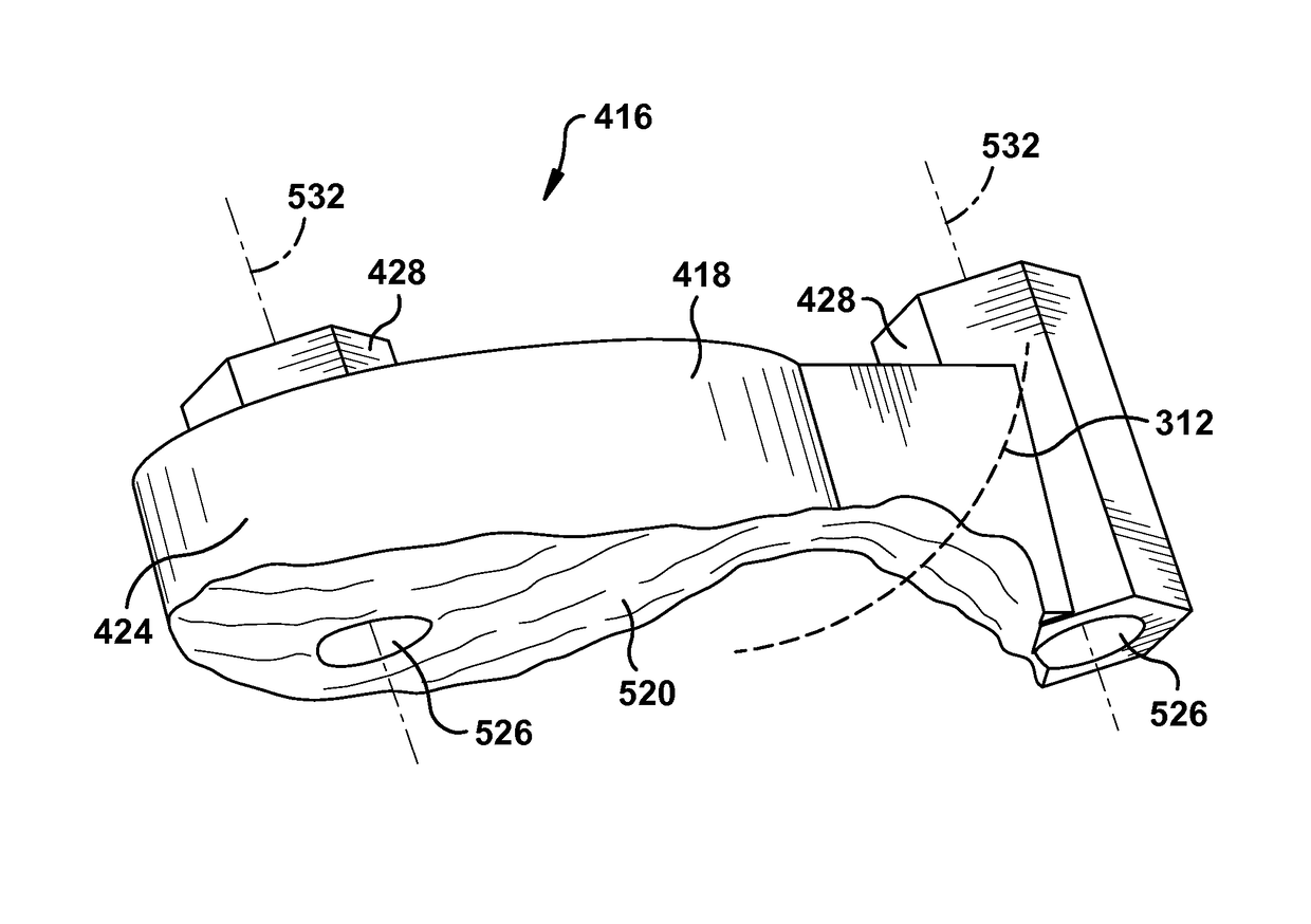
[0085]In accordance with the present invention, FIG. 17 depicts a guide 416′ including a base 418′, a stem 1758, and at least one spacing arm 1760. The base 418′ has a lower base surface 520′ (shown partially in phantom line in FIG. 17) spaced apart from an upper base surface 422′ by a base body 424′. The lower base surface 520′ is contoured to mate with the acetab...

first embodiment

[0089]The guide 416′ may be at least partially customized responsive to preoperative imaging of the patient tissue. For example, the lower base surface 520′ of the base 418′ could be at least partially configured through the use of computer tomography (“CT”) data of the patient tissue to have a longitudinally downward-protruding portion corresponding to the acetabular fossa 1656. Additionally or alternatively, the lower base surface 520′ could be at least partially configured through use of patient scans including digital or analog radiography, magnetic resonance imaging, or any other suitable imaging means. The patient tissue preoperative images are optionally displayed for review and manipulation before / during configuration of the lower base surface 520′, such as through the use of a computer or other graphical workstation interface described above with reference to the present invention. The configuration of the lower base surface 520′ is described herein as being performed using...

third embodiment

[0112]FIG. 29 depicts a third configuration of a guide 416″ according to the present invention. In FIG. 29, the guide 416″ is configured to assist with correction of a malunion or other deformity in the head of a femur, humerus, tibia, phalange, mandible, scapula, or any other suitable bone or other patient tissue. The guide 416″ of the third configuration 416″ can be used similarly to the guides 416″ of the first and second configurations.

[0113]FIGS. 30-39 depict a guide 416′ according to certain additional aspects of the second embodiment of the present invention. The guide 416′ of FIGS. 30-37 is similar to the guide 416 of FIGS. 1-15 and the guide 416′ of FIGS. 16-24 and therefore, structures of FIGS. 30-39 that are the same as or similar to those described with reference to FIGS. 1-15 and / or 16-24 have the same reference numbers with the addition of a “prime” mark. Description of common elements and operation similar to those in the previously described first embodiment will not...


PUM


Abstract

A patient tissue includes a primary patient tissue area and an anatomically differentiated bordering secondary patient tissue area. An apparatus is at least partially customized responsive to preoperative imaging of the patient tissue. Means are provided for mating with the primary patient tissue area in a preselected relative orientation. Means are provided for fixing a first landmark to the primary patient tissue area in at least one of a predetermined marking location and a predetermined marking trajectory. Means are provided for fixing a second landmark to the secondary patient tissue area in at least one of a predetermined marking location and a predetermined marking trajectory. A method of associating a plurality of landmarks with a patient tissue is also provided.

Description

RELATED APPLICATION[0001]This application claims priority from U.S. Provisional Application No. 61 / 408,359, filed Oct. 29, 2010, the subject matter of which is incorporated herein by reference in its entirety.TECHNICAL FIELD[0002]The present invention relates to a system and method for association of a guiding aid with a patient tissue and, more particularly, to a system and method for associating at least one landmark with the patient tissue for assisting with attachment of a stock prosthetic implant to the patient tissue.BACKGROUND OF THE INVENTION[0003]In the installation of a prosthetic shoulder joint into a patient's body, a glenoid component is implanted into the glenoid vault of the patient's scapula. An obverse surface of the glenoid component is configured for articulating contact with a humeral component carried by the patient's humerus. A reverse surface of the glenoid component is secured to the bone surface of the glenoid vault.[0004]Because the shoulder prosthesis is n...

Claims


Application Information

Patent Timeline
Patent Type & Authority Patents(United States)
IPC IPC(8): A61B17/56A61B17/17A61B17/15A61B34/10
CPCA61B17/1746A61B34/10A61B17/152A61B2017/568A61B2034/104A61B2034/105A61B2034/108A61B17/1778A61B17/151
Inventor IANNOTTI, JOSEPH P.BARSOUM, WAEL K.BRYAN, JASON A.O'NEILL, PETER D.
Owner THE CLEVELAND CLINIC FOUND
Why Eureka
Try Eureka