
Inhibition of pulmonary fibrosis with nutlin-3A and peptides

a technology of nutlin-3a and pulmonary fibrosis, which is applied in the field of biochemistry and medicine, can solve the problems of reducing the viability of ecm, restricting the production and deposition of ecm,

Active Publication Date: 2017-04-25
BOARD OF RGT THE UNIV OF TEXAS SYST
View PDF21 Cites 23 Cited by
  • Summary
  • Abstract
  • Description
  • Claims
  • Application Information

AI Technical Summary

Benefits of technology

The patent describes a new compound (NTL) that inhibits the interaction between a protein called p53 and another called MDM2, which is involved in cell death and proliferation. NTL was able to increase p53 and decrease uPA, which is involved in fibrotic lung disease. This resulted in the death of fibrotic fibroblasts and the inhibition of their growth. The patent proposes a new treatment for lung fibrosis that targets these specific cells and their interactions with other proteins in the body.

Problems solved by technology

Restoration of p53 expression and p53-mediated changes in the uPA-fibrinolytic system in FL-fibroblasts leads to attenuation of viability, and restricts production and deposition of ECM.

Method used


Image

  • Inhibition of pulmonary fibrosis with nutlin-3A and peptides
  • Inhibition of pulmonary fibrosis with nutlin-3A and peptides
  • Inhibition of pulmonary fibrosis with nutlin-3A and peptides

Examples


example i

Deceased FL-Fibroblast p53 Expression in Fibrotic Foci of IPF Lungs

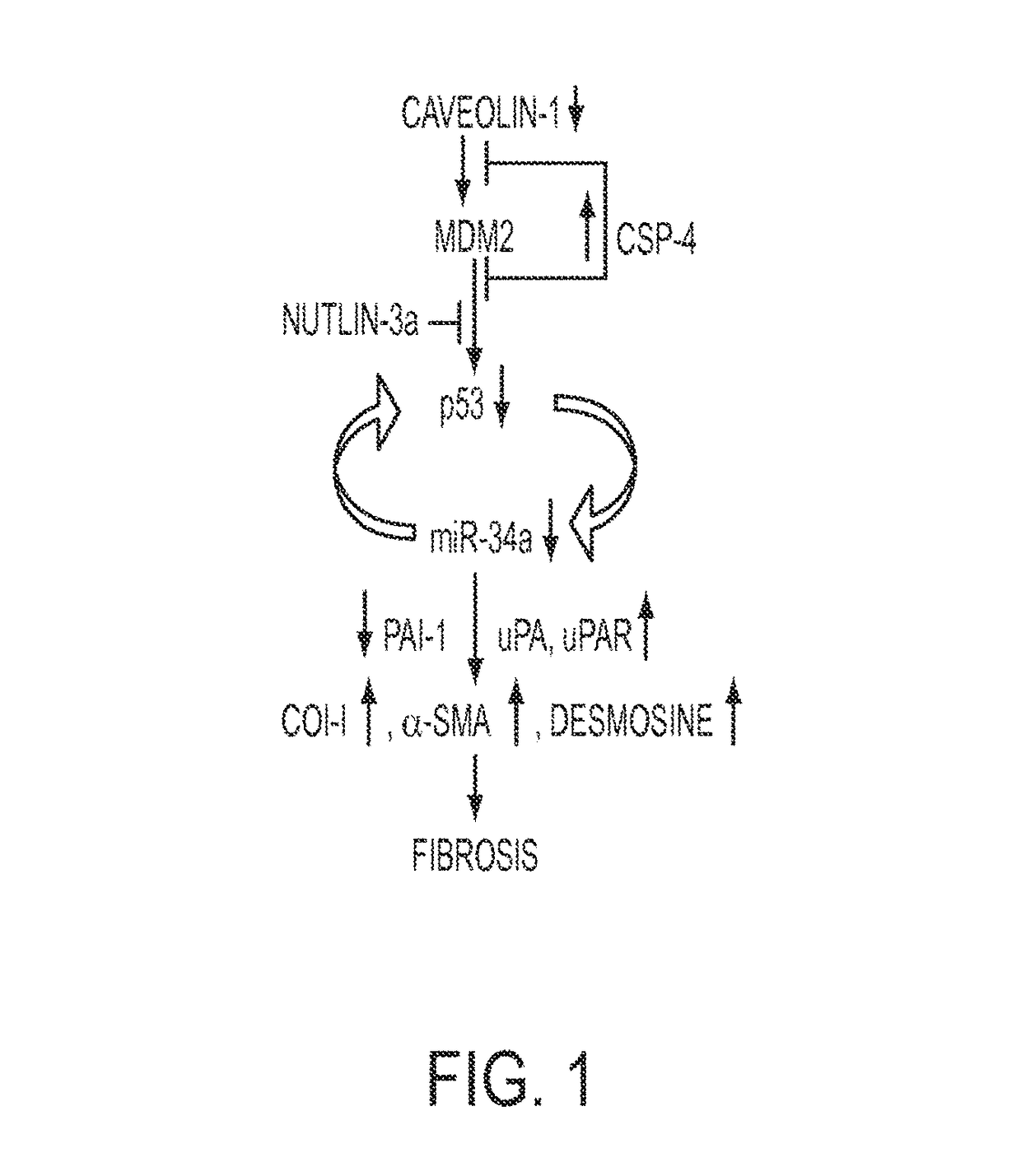
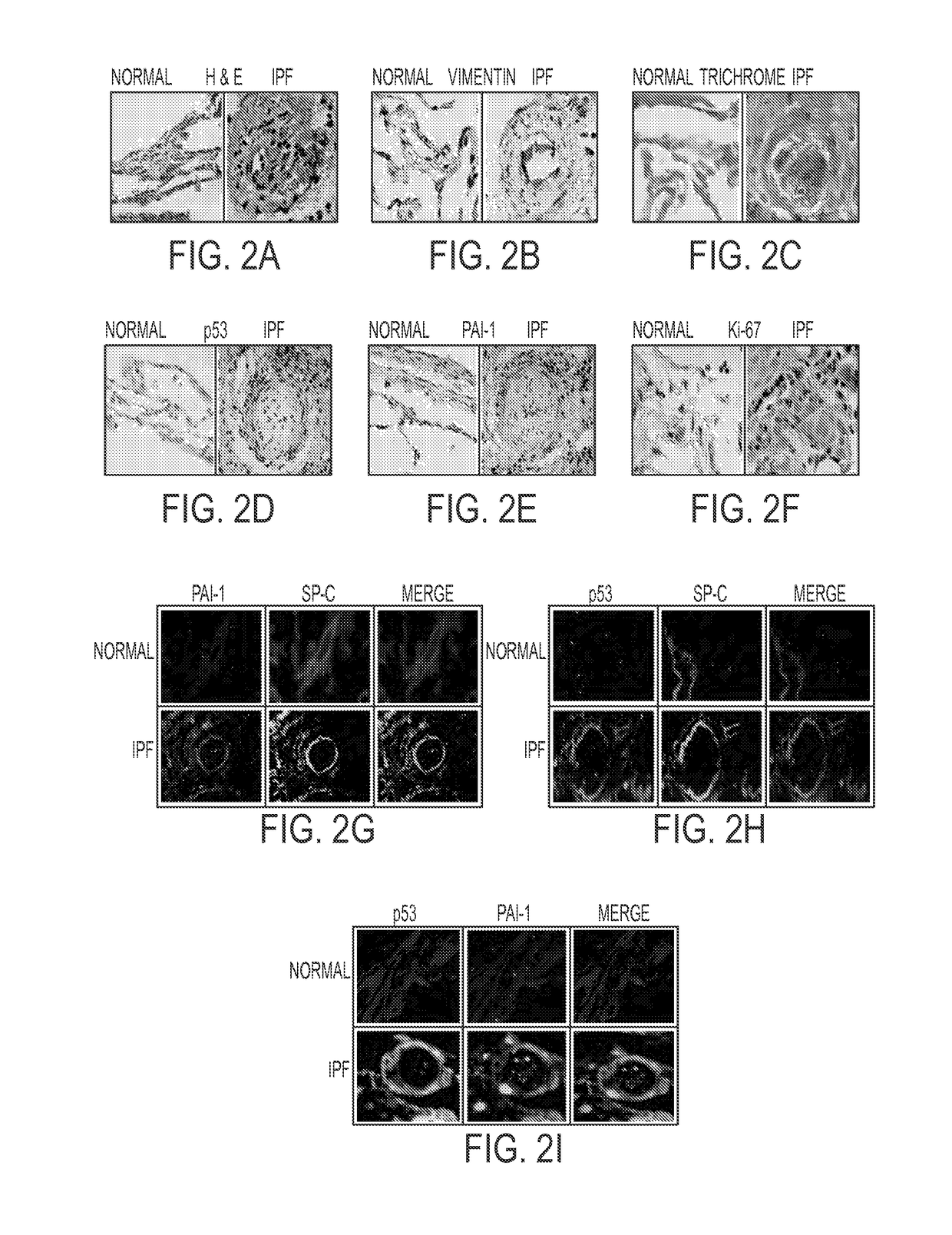
[0250]Staining of lung sections from IPF patients and control “normal” subjects with various reagents, including by immunohistochemistry (IHC) (see FIGS. 2A-2I) revealed that IPF tissues display fibrotic foci which are dense with ECM. FL-fibroblasts dispersed in the vimentin-rich foci showed minimal staining for p53 and PAI-1 antigens. However, immunofluorescence staining for SP-C, PAI-1, p53 and active caspase-3 (not shown) demonstrated that alveolar type II (ATII) cells surrounding the fibrotic foci show elevated staining for p53 and PAI-1 antigens, and are positive for active caspase-3, indicating apoptosis of encircling ATII cells. The IHC results reveal that ATII cells encasing fibrotic foci continuously die due to increased expression of p53 and PAI-1. These wounds are replaced by activated fibroblasts which show minimal basal p53 and PAI-1, and elevated ki-67 staining indicating proliferation due to suppressio...

example ii

Decreased Caveolin-1, p53 and PAI and Increased uPA in Human FL Fibroblasts from IPF Lungs

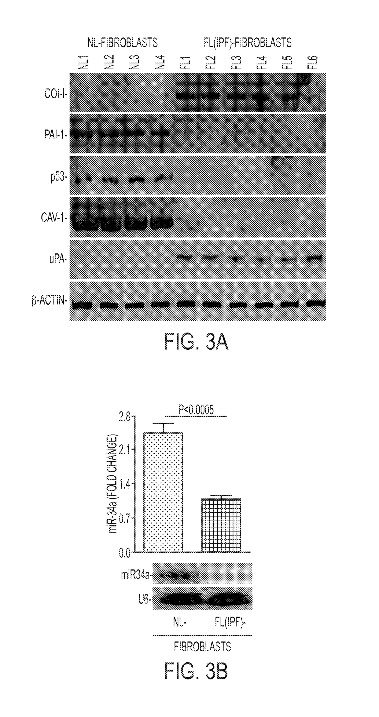
[0251]Cell lysates from NL- and FL-fibroblasts were immunoblotted to reveal changes in the proteins and a miRNA. See FIG. 3A-3B. Basal miR-34a expression was significantly lower in FL-fibroblasts indicating that reduced p53 expression and consequent changes in p53-uPA fibrinolytic system cross-talk contributed to fibrogenesis. Such changes are associated with increased col-I and inhibition of miR-34a. The results further showed that increases in p53-induced miR-34a transcription or stabilization of p53 in human FL-fibroblasts mediated by miR-34a can mitigate lung fibrosis.

example iii

Disparate Expression of uPA, PAI-1 and Col-I mRNAs by Fibroblasts from IPF and “Normal” Lungs

[0252]Total RNA isolated from lung tissues from “normal” subjects and patients with IPF or from NL-were tested for uPA, PAI-1 and col-I mRNA by quantitative RT-PCR and normalized to the corresponding levels of β-actin mRNA. Results are shown in FIGS. 4A-4B.

[0253]Expression of col-I and PAI-1 mRNA significantly increased in IPF lung tissues whereas uPA mRNA was is reduced. Interestingly, unlike IPF lung tissues, col-I and uPA mRNA and lower level of PAI-1 mRNA expression were found in FL-fibroblasts compared to NL-fibroblasts. uPAR protein and mRNA are also elevated in FL-fibroblasts. The elevated PAI-1 in the lung tissues is attributable to increased expression of PAI-1 by lung epithelial cells or macrophages rather than FL(IPF)-fibroblasts. This is consistent with increased expression of col-I, uPA and uPAR, and reduced PAI-1 proteins in FL-fibroblasts from IPF lungs (see FIG. 3A).


PUM

PropertyMeasurementUnit
timeaaaaaaaaaa
temporal heterogeneityaaaaaaaaaa
compositionsaaaaaaaaaa

Abstract

In fibrotic lung fibroblasts, basal levels of p53 protein (and miR-34a) are markedly suppressed, leading to reduced p53-mediated inhibition of uPA and uPAR, or concurrent induction of PAI-1. These changes contribute to excessive FL-fibroblast proliferation and production of extracellular matrix (ECM), and, therefore, pulmonary fibrosis. These processes are reversed by treating the cells, and treating subjects suffering from idiopathic pulmonary fibrosis (IPF) with the small organic molecule nutlin-3a (NTL) or with a peptide, CSP-4 (SEQ ID NO:1), or variants or derivatives or multimers of this peptide, which increase p53 levels by inhibiting MDM2-mediated degradation of p53 protein. Use of these compounds serves as a new approach to the treatment of IPF, they restore p53 expression and p53-mediated changes in the uPA-fibrinolytic system in FL-fibroblasts and restrict production and deposition of ECM.

Description

[0001]This application is a national phase application under 35 U.S.C. §371 of International Application No. PCT / US2014 / 030147, filed Mar. 17, 2014, which claims the benefit of U.S. Provisional Patent Application No. 61 / 800,117, filed Mar. 15, 2013.BACKGROUND OF THE INVENTION[0002]Field of the Invention[0003]The present invention in the field of biochemistry and medicine is directed to methods and composition for increasing p53 protein levels, reducing uPA and uPAR and increasing PAI-1 expression, in fibrotic lung (FL) fibroblasts and reducing their proliferation, and for treating idiopathic pulmonary fibrosis (IPF) using nutlin-3a and peptides.[0004]Description of the Background Art[0005]Idiopathic pulmonary fibrosis (IPF) is a poorly understood progressive and fatal lung disease for which no treatment exists other than lung transplantation (Mason D P et al., Ann Thorac Surg 84:1121-8, 2007). Median survival of five years after diagnosis is less than 20%. Most forms of interstitial...

Claims


Application Information

Patent Timeline
Patent Type & Authority Patents(United States)
IPC IPC(8): A61K38/08C07K7/06C07K7/08A61K9/00A61K31/497A61K38/17A61K38/00
CPCC07K7/06A61K9/007A61K9/0019A61K31/497A61K38/08A61K38/177C07K7/08A61K38/00A61K2300/00A61P11/00A61P43/00A61K47/64C07K2319/10A61K47/645
Inventor SHETTY, SREERAMAIDELL, STEVEN
Owner BOARD OF RGT THE UNIV OF TEXAS SYST
Learn More