
Compositions and methods for promoting patency of vascular grafts

a vascular graft and patency technology, applied in the field of compositions and methods for increasing the patency of vascular grafts, can solve the problems of increased contamination risk, increased risk of contamination, and increased difficulty in obtaining healthy autologous cells from diseased donors, and achieve the effect of increasing patency

Active Publication Date: 2018-01-02
YALE UNIV
View PDF18 Cites 2 Cited by
  • Summary
  • Abstract
  • Description
  • Claims
  • Application Information

AI Technical Summary

Benefits of technology

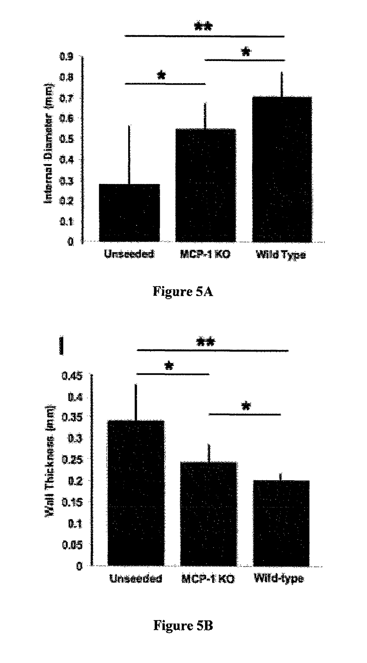
[0014]Methods for increasing the patency of biodegradable, synthetic vascular grafts are provided. The methods include administering one or more cytokines and / or chemokines that promote outward tissue remodeling of the vascular grafts and vascular neotissue formation. The polymeric vascular grafts are tubular, porous structures that allow for recruitment and integration of host cells into the graft that mediate remodeling and vascular neotissue formation. The vascular grafts are biodegradable, which allows for the grafts to be completely replaced by forming neotissue as they degrade. The methods do not require cell seeding of the vascular grafts, avoiding many problems associated with seeding, including the need for an invasive procedure to obtain autologous cells, the need for a substantial period of time in order to expand the cells in culture, the inherent difficulty in obtaining healthy autologous cells from diseased donors, and an increased risk of contamination and the potential for dedifferentiation of the cells. The disclosed biodegradable, synthetic vascular grafts therefore have a greater off-the-shelf availability and increased overall clinical utility.

Problems solved by technology

The methods do not require cell seeding of the vascular grafts, avoiding many problems associated with seeding, including the need for an invasive procedure to obtain autologous cells, the need for a substantial period of time in order to expand the cells in culture, the inherent difficulty in obtaining healthy autologous cells from diseased donors, and an increased risk of contamination and the potential for dedifferentiation of the cells.

Method used


Image

  • Compositions and methods for promoting patency of vascular grafts
  • Compositions and methods for promoting patency of vascular grafts
  • Compositions and methods for promoting patency of vascular grafts

Examples


example 1

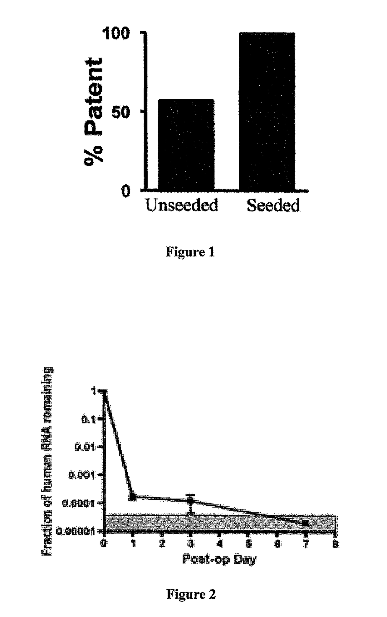
man Bone Marrow Mononuclear Cells (hBMCs) Improve the Functional Outcome and Vascular Development of TEVG

[0085]Materials and Methods:

[0086]Biodegradable PGA-P(CL / LA) Scaffold Construction

[0087]A dual cylinder chamber molding system was constructed from a 6.5 mm diameter polypropylene rod. A 1.4 mm diameter inner cylinder was cored through the center of the rod for a length of 30 mm and gradually tapered out to 6.0 mm at the inlet. Polyglycolic acid (PGA) nonwoven felt (ConcordiaFibers, Coventry, R.I.) was used for the framework of the scaffold. Felts were 300 mm thick with approximately 90% (PGA) or 83% (PLLA) total porosity. The gradual taper of the inner cylinder enabled flat felt sections (6.0 mm×4.0 mm) to be easily shaped into tubes during their insertion through the inlet of the dual cylinder chamber system. Stainless steel needles (21 g) were then introduced into the opposing end to maintain the inner lumen and further compress the felt. A 50:50 copolymer sealant solution of ...

example 2

s Constitute a Minor Fraction of Seeded hBMCs

[0105]Materials and Methods:

[0106]Materials and methods were as described in Example 1 except as described below.

[0107]Characterization of hBMCs

[0108]Flow cytometry was used to identify and quantify sub-populations in the mononuclear cell fraction of human bone marrow. Human BMC from five different donors were used and tested in triplicate. Antibodies used were purchased from BD Biosciences CD8 PerCP(SK1), CD14 FITC (M5E2), CD31 FITC (WM59), CD34 PE and PerCP-Cy5.5 (8G12), CD45 APC-Cy7 (2D1), CD56 PE (NCAM16.2), CD146 (P1H12), CD73 PE (AD2), CD90 FITC (5E10), 7AAD; E-Bioscience CD3 APC (HIT3a), CD4 FITC (L3T4), CD19 (PE-Cy7); Miltenyi Biotec CD133 APC (AC133); R&D Systems VEGFR2PE (89106); and AbD Serotec CD105 Alexa 647 (SN6). Cells were acquired on a FACSAria cell sorter and results analyzed using DIVA software (BD Bioscience).

[0109]Quantitative Image Analysis

[0110]Relative cellularity was measured for each explanted scaffold. Two separ...

example 3

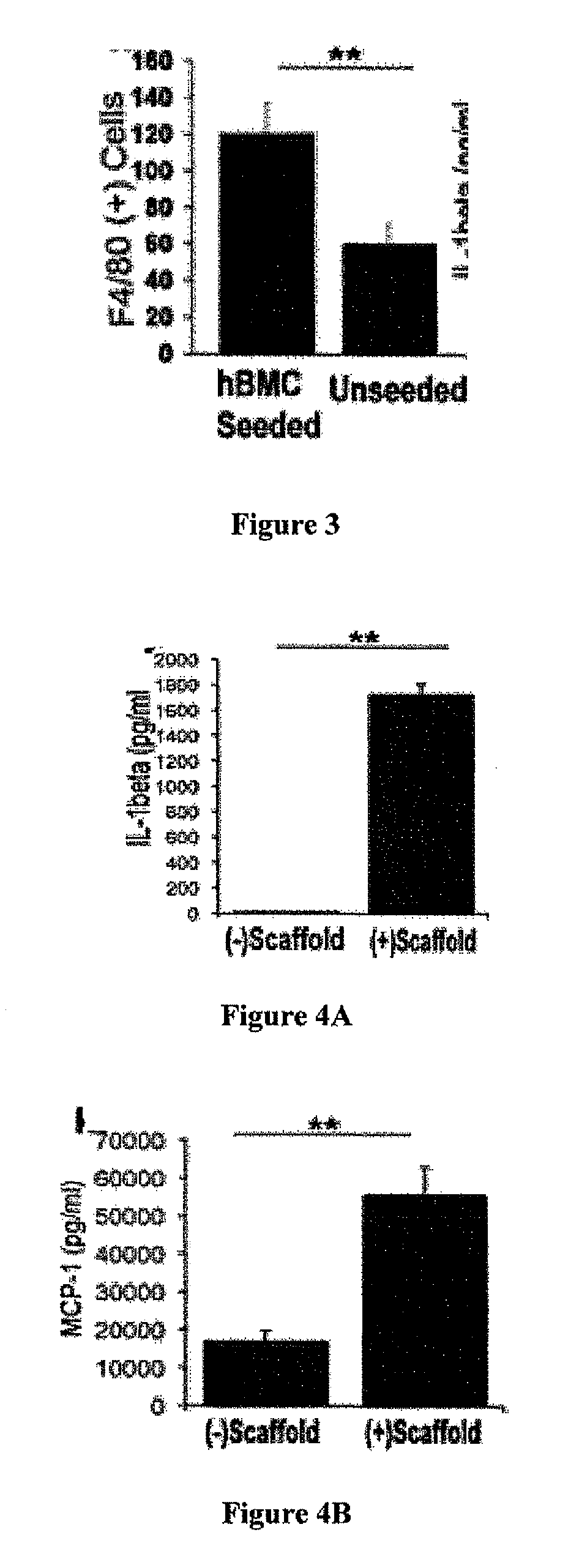
MCs are not Incorporated into the Developing Neovessel

[0116]Materials and Methods:

[0117]Materials and methods were as described in Examples 1 and 2 except as described below.

[0118]Immunohistochemistry

[0119]Species-specific antibodies were used to distinguish between seeded human cells and recruited murine host cells. Human-specific primary antibodies used included mouse-anti-human CD31 (Dako), CD68 (Dako), CD34 (Abeam), and CD45 (AbD Serotec). Mouse-specific primary antibodies used included rat-anti-mouse Mac-3 (BD Bioscience), F4 / 80 (AbD Serotec), IL-1beta (R&D Systems), and goat-anti-mouse VEGF-R2 (R&D Systems, minimal cross-reactivity). Primary antibodies that cross-reacted with both species were mouse-anti-human α-smooth muscle actin (αSMA) (Dako), VEGF (Santa Cruz), and rabbit-anti-human von Willebrand Factor (vWF) (Dako). Antibody binding was detected using appropriate biotinylated secondary antibodies, followed by binding of streptavidin-HRP and color development with 3,3-dia...


PUM

PropertyMeasurementUnit
diameteraaaaaaaaaa
diameteraaaaaaaaaa
sizeaaaaaaaaaa

Abstract

Methods for increasing the patency of biodegradable, synthetic vascular grafts are provided. The methods include administering one or more cytokines and / or chemokines that promote outward tissue remodeling of the vascular grafts and vascular neotissue formation. The disclosed methods do not require cell seeding of the vascular grafts, thus avoiding many problems associated with cell seeding. Biodegradable, polymeric vascular grafts which provide controlled release of cytokines and / or chemokines at the site of vascular graft implantation are also provided.

Description

CROSS REFERENCE TO RELATED APPLICATIONS[0001]This application claims priority to and benefit of U.S. Provisional Patent Application No. 61 / 010,406 filed on Jan. 8, 2008, by Christopher K. Breuer, Themis R. Kyriakides and Jason D. Roh, and where permissible is incorporated by reference in its entirety.GOVERNMENT SUPPORT[0002]This invention was made with government support under grant number 5K08HL083980 awarded by the National Institutes of Health. The government has certain rights in the invention.FIELD OF THE INVENTION[0003]This invention relates generally to compositions and methods for increasing the patency of vascular grafts, in particular vascular grafts made from biodegradable, polymeric scaffolds.BACKGROUND OF THE INVENTION[0004]Cardiac defects are the most common congenital anomalies affecting nearly 1% of all live births. Despite major advances in the treatment of congenital heart disease (CHD), it remains the leading cause of death due to congenital anomalies in the newbo...

Claims


Application Information

Patent Timeline
Patent Type & Authority Patents(United States)
IPC IPC(8): A61L27/50A61L27/54A61K38/19A61L27/18A61F2/06
CPCA61L27/507A61L27/18A61L27/54C08L67/04A61F2/06A61K38/195A61L2300/604A61L2300/426A61P43/00
Inventor BREUER, CHRISTOPHERKYRIAKIDES, THEMISROH, JASON
Owner YALE UNIV
Learn More