Detecting coronary stenosis through spatio-temporal tracking

a spatio-temporal tracking and coronary artery technology, applied in the field of automatic angiogram analysis, can solve problems such as chest symptoms, permanent impairment of heart muscle function, and heart attack

Inactive Publication Date: 2018-04-17
INT BUSINESS MASCH CORP
View PDF12 Cites 3 Cited by
  • Summary
  • Abstract
  • Description
  • Claims
  • Application Information

AI Technical Summary

Problems solved by technology

Any occlusion of these arteries can impact heart function leading to effort intolerance with exertion provoked chest symptoms, heart attack, permanent impairment of heart muscle function, and sudden death.
In general detection of vessels and coronary stenosis in X-ray angiography data is difficult due to the low contrast between vessels and surrounding tissue, as well as large intensity gradients within the image.
In addition, the curvilinear cylindrical structure of an artery and the irregular and often eccentric remaining lumen through the diseased segment make accurate visual detection of stenosis difficult.
Often, spurious dye and imaging artifacts can give a false appearance of stenosis in these sequences.

Method used


Image

  • Detecting coronary stenosis through spatio-temporal tracking
  • Detecting coronary stenosis through spatio-temporal tracking
  • Detecting coronary stenosis through spatio-temporal tracking

Examples


Embodiment Construction

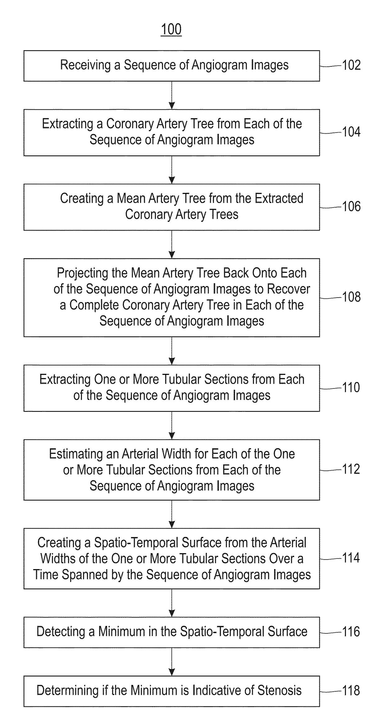
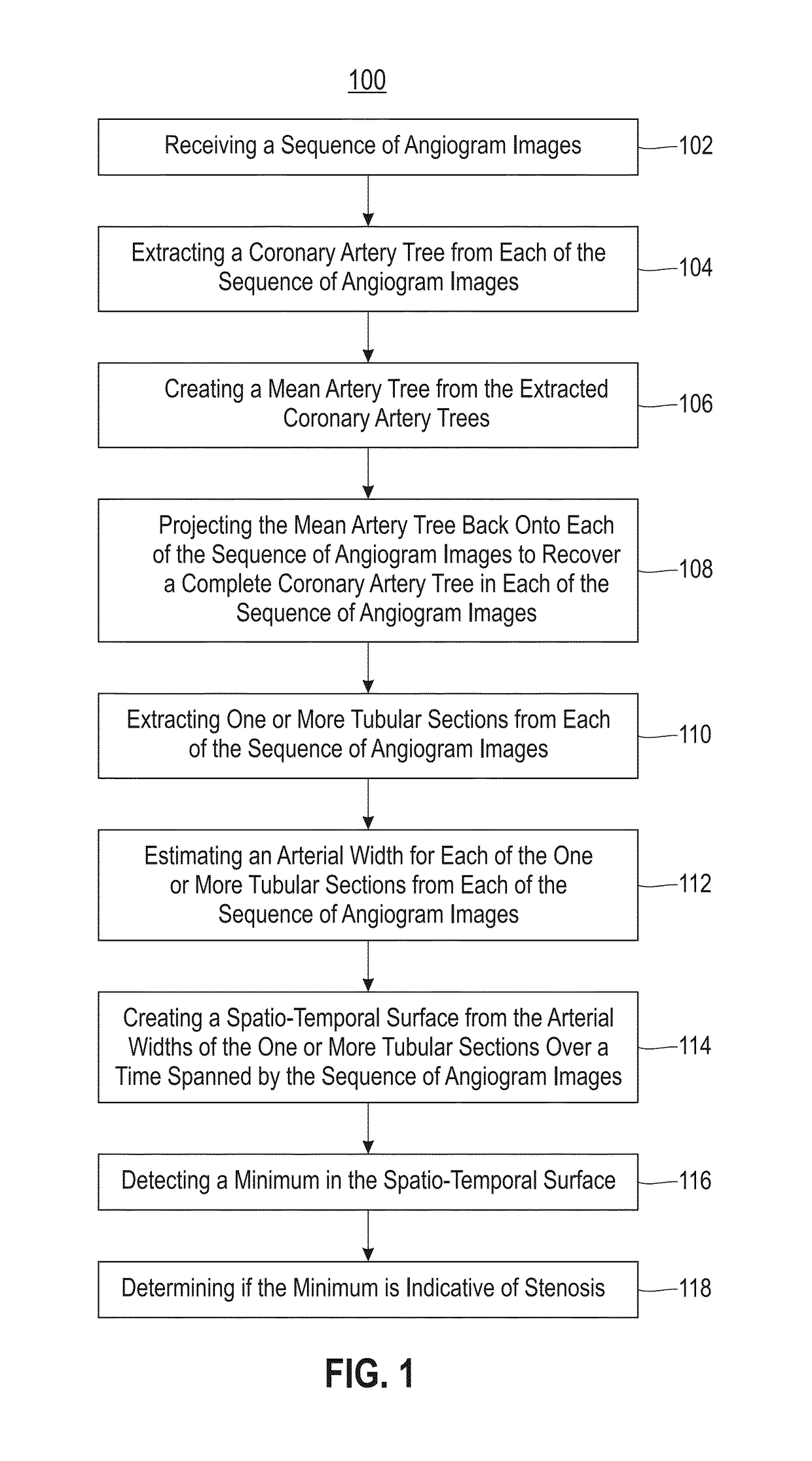
[0020]Embodiments described herein are directed to methods, systems and computer program products for automatic detection of coronary stenosis in X-ray angiography data. In exemplary embodiments, the spatio-temporal nature of angiography sequences is used to isolate the coronary artery tree. An arterial width surface is formed for each isolated artery segment by calculating the width along a segment and tracking the segment in each image frame over time. A persistent minima of this surface, which corresponds to a stenosis in the artery, is then identified.

[0021]Referring now to FIG. 1, a process flow of a method 100 for detecting coronary stenosis through spatio-temporal tracking in accordance with an embodiment is illustrated. As illustrated at block 102, the method 100 includes receiving a sequence of angiogram images. In exemplary embodiments, the sequence of angiogram images are X-ray angiogram images. Next, as shown at block 104, the method 100 includes extracting a coronary ar...


PUM


Abstract

Embodiments relate to detecting coronary stenosis through spatio-temporal tracking. Aspects include extracting a coronary artery tree from each of a sequence of angiogram images, creating a mean artery tree from the extracted coronary artery trees, and projecting the mean artery tree back onto each of the sequence of angiogram images to recover a complete coronary artery tree for each of the sequence of angiogram images. Aspects also include extracting one or more tubular sections from each of the sequence of angiogram images, estimating an arterial width for each of the one or more tubular sections from each of the sequence of angiogram images, and creating a spatio-temporal surface from the arterial widths of the one or more tubular sections over a time spanned by the sequence of angiogram images. Aspects further include detecting a minimum in the spatio-temporal surface and determining if the minimum is indicative of stenosis.

Description

BACKGROUND[0001]Embodiments of the invention relate generally to automated angiogram analysis, and more specifically, to detecting coronary stenosis through spatio-temporal tracking.[0002]Cardiovascular disease (CVD) results in more death and disability in both males and females in all western societies than any other disease category, accounting for one third of all deaths in the United States in 2007. Any occlusion of these arteries can impact heart function leading to effort intolerance with exertion provoked chest symptoms, heart attack, permanent impairment of heart muscle function, and sudden death.[0003]The “gold standard” diagnostic test for CAD is the coronary angiogram. A coronary angiogram involves placing plastic catheters into the arterial system and injecting iodinated contrast solution into the coronary blood flow, to obtain a silhouette of the coronary arterial wall. The angiographic images are typically recorded at 15 or 30 frames a second, providing a motion pictur...

Claims


Application Information

Patent Timeline
Patent Type & Authority Patents(United States)
IPC IPC(8): G06K9/46A61B5/00A61B6/00G06T7/00G06T7/174
CPCA61B6/481A61B6/504G06T7/0016G06T7/174G06T2207/30101A61B6/5217G06T2207/10016G06T2207/10116
Inventor COMPAS, COLIN B.SYEDA-MAHMOOD, TANVEER F.
Owner INT BUSINESS MASCH CORP
Why Eureka
Try Eureka