Aortic stenosis detection device

A detection device and aortic technology, applied in the field of medical detection, can solve the problems of insufficient detection and achieve the effect of reducing false and missed detection

Inactive Publication Date: 2020-12-29
樊开凯
View PDF0 Cites 0 Cited by
  • Summary
  • Abstract
  • Description
  • Claims
  • Application Information

AI Technical Summary

Problems solved by technology

However, the current detection methods are all detected by an X-ray source, and it is inevitable that the detection is not in place. How t

Method used


Image

  • Aortic stenosis detection device
  • Aortic stenosis detection device
  • Aortic stenosis detection device

Examples


Embodiment Construction

[0016] The application will be described in further detail below in conjunction with the accompanying drawings. It is necessary to point out that the following specific embodiments are only used to further illustrate the application, and cannot be interpreted as limiting the protection scope of the application. The above application content makes some non-essential improvements and adjustments to this application.

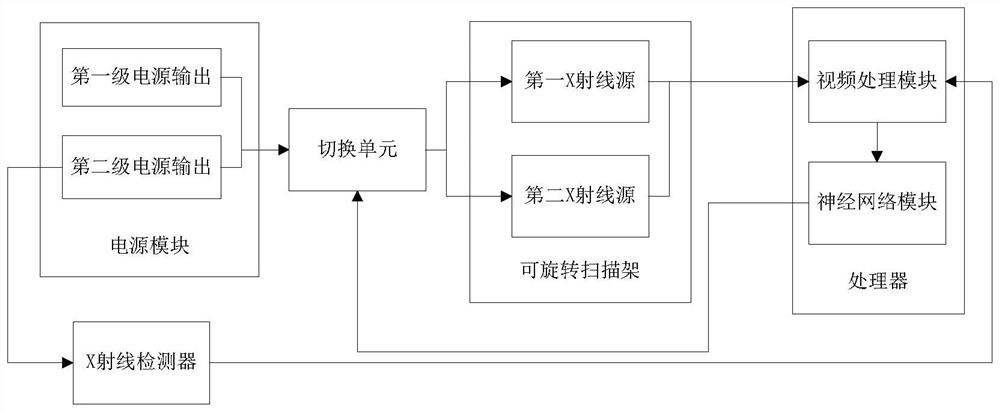
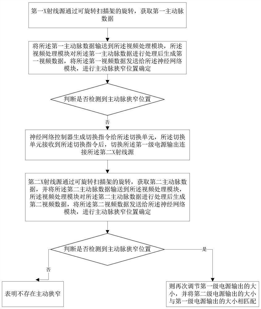
[0017] Such as figure 1 Shown is a schematic diagram of an aortic stenosis detection device of the present invention. The invention discloses an aortic stenosis detection device, comprising: a rotatable scanning frame, a first X-ray source, a second X-ray source, an X-ray detector, a power supply module and a processor, the first X-ray source, the second X-ray source Two X-ray sources are arranged on the rotatable gantry and project X-rays toward the aorta, and the X-ray detector is arranged on the rotatable gantry and arranged to receive the first X-ray source, t...


PUM


Abstract

The invention relates to an aortic stenosis detection device. The device comprises a rotatable scanning frame, a first X-ray source, a second X-ray source, an X-ray detector, a power supply module anda processor, wherein the first X-ray source and the second X-ray source emit X-rays; the output of a first-stage power supply is used for supplying power to the first X-ray source or the second X-raysource; a second-stage power supply is used for supplying power to the X-ray detector; the processor comprises a video processing module and a neural network module; the video processing module is connected with the first X-ray source and the second X-ray source and used for receiving aorta data detected by the first X-ray source and the second X-ray source and processing the detected data to generate a visual video; the video processing module is used for sending the visual video to the neural network module; and the neural network module is used for training video data to obtain the specific position of aortic stenosis.

Description

technical field [0001] The invention belongs to the technical field of medical detection, in particular to an aortic stenosis detection device. Background technique [0002] Coronary artery disease is one of the diseases with the highest mortality rate in the world, and aortic stenosis is a major form of arterial disease. Digital silhouette angiography (DSA) and CT are currently the main methods that can diagnose the degree of cardiac artery disease. Generally, doctors send a catheter into the coronary sinus of the heart, and then release a contrast agent through the catheter to visualize the coronary arteries. After that, the doctor observes according to different body positions and finally confirms the location of the lesion. However, the current detection methods are all detected by an X-ray source, and it is inevitable that the detection is not in place. How to quickly realize the detection of aortic stenosis and achieve fast and accurate detection through a single dete...

Claims


Application Information

Patent Timeline
IPC IPC(8): A61B6/00
CPCA61B6/504A61B6/54
Inventor 樊开凯
Owner 樊开凯
Why Eureka
Try Eureka