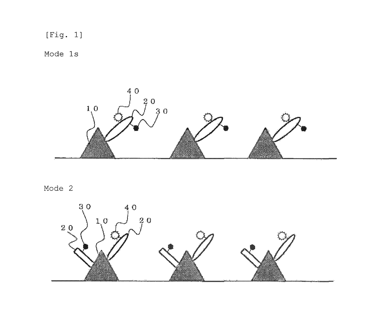
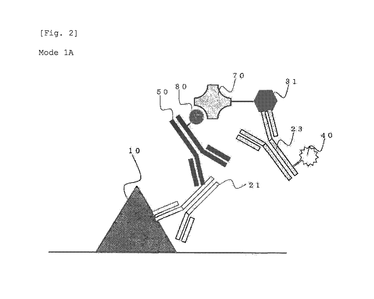
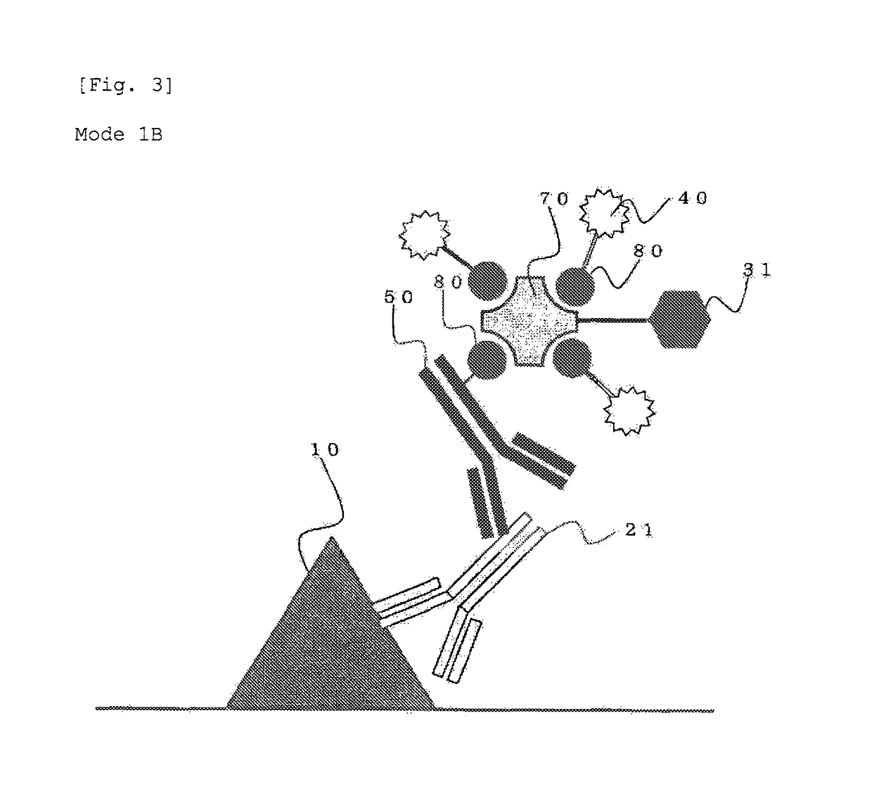
Method for staining tissue

a tissue and staining technology, applied in the field of tissue staining, can solve the problems of inability to predict more right and detailed pathology judgment based on the experience of skilled pathologists, change in stainability, and inability to quantify the criteria of staining, etc., and achieve high quantitativity and high precision

Active Publication Date: 2018-05-15
KONICA MINOLTA INC
View PDF9 Cites 2 Cited by
  • Summary
  • Abstract
  • Description
  • Claims
  • Application Information

AI Technical Summary

Benefits of technology

[0017]According to the present invention, information about the expression amount, the intracellular localization, and so on of a specific biological substance on a tissue section can be acquired with a high quantitativity and in detail. In addition, double check by both visual evaluation and digital evaluation using the same section can be carried out. Consequently a method which can cover variation in diagnosis by the level of skill of pathologists and enables right judgment on the results can be provided. Specifically, for example, a system in which, after the visual evaluation by physicians, automatic analysis based on bright spots of fluorescence is automatically carried out by a virtual slide system or the like to extract samples whose results therefrom are different from the evaluation by the physicians and propose reevaluation to the physicians can be exemplified. Moreover, a method for judging with a high precision the effectiveness of a pharmaceutical containing an antibody as an ingredient can be provided.

Problems solved by technology

However, there is a problem with the quantitativity of its results, since, in a staining method using an enzyme reaction like the DAB method, the stainability is changed depending on the staining conditions including the reaction time (see, for example, Non-patent Documents 1 and 2).
In addition, its criteria lack quantitativity because of their roughness since the staining level is graded in only 4 scores of from 0 to 3, and further the judgment using the criteria depends on the level of skill of the pathologist or the like, which have been problematic.
Accordingly, there is a problem that, in the case of using only the information obtained by visualization using a light-emitting substance such as a phosphor, quantitativity can be expected but the more right and detailed pathology judgment based on the experiences of the skilled pathologists can not be expected.
However, the FISH method is a quantitative examination method, but is not a method by which the expression amount and / or the intracellular localization of a protein are directly evaluated, which has been problematic.

Method used


Image

  • Method for staining tissue
  • Method for staining tissue
  • Method for staining tissue

Examples


synthesis example 1

FITC-Aggregated Silica Nanoparticle

[0138]In N,N-dimethylformamide (DMF), 6.6 mg of amino reactive FITC (manufactured by DOJINDO LABORATORIES) and 3 μL of 3-aminopropyltrimethoxysilane (manufactured by Shin-Etsu Silicone: KBM903) were mixed to obtain an organoalkoxysilane compound.

[0139]Then, 0.6 mL of the obtained organoalkoxysilane compound was mixed with 48 mL of ethanol, 0.6 mL of tetraethoxysilane (TEOS), 2 mL of water, and 2 mL of 28% aqueous ammonia for 3 hours.

[0140]The mixture produced in the above-described step was centrifuged at 10,000 G for 20 minutes, and the supernatant was removed. Ethanol was added thereto to disperse the precipitate, and centrifugation was carried out again. Further, by the same procedure, washing was carried out twice with ethanol and twice with pure water. As a result, 10 mg of an FITC-aggregated silica nanoparticle was obtained as a fluorescent aggregate.

[0141]By performing SEM observation for 1000 of the obtained FITC-aggregated silica nanoparti...

synthesis example 2

TXR-Aggregated Silica Nanoparticle

[0142]Using Texas Red (manufactured by DOJINDO LABORATORIES, hereinafter referred to as “TXR”) instead of amino reactive FITC in Synthesis Example 1, the same synthesis was carried out, and 10 mg of a TXR-aggregated silica nanoparticle was obtained. By performing SEM observation for 1000 of the obtained TXR-aggregated silica nanoparticle, it was found that the average particle size was 106 nm and the coefficient of variation was 11%.

synthesis example 3

Semiconductor Nanoparticle (Qdot605)-Aggregated Silica Nanoparticle

[0143]To 10 μL of a CdSe / ZnS decane dispersion (“Qdot605” (registered trademark), manufactured by Invitrogen Corporation), 0.1 mg of TEOS, 0.01 mL of ethanol, 0.03 mL of concentrated aqueous ammonia were added, and the mixture was stirred for 3 hours to carry out hydrolysis.

[0144]The mixture obtained in the above-described step was centrifuged at 10,000 G for 20 minutes, and the supernatant was removed. Ethanol was added thereto to disperse the precipitate, and centrifugation was carried out again. Further, by the same procedure, washing was carried out twice with ethanol and twice with pure water. As a result, 40 mg of a Qdot605-aggregated silica nanoparticle was obtained as a fluorescent aggregate.

[0145]By performing SEM observation for 1000 of the obtained Qdot605-aggregated silica nanoparticle, it was found that the average particle size was 108 nm and the coefficient of variation was 14%.

Production of Biotinylat...


PUM

PropertyMeasurementUnit
particle sizeaaaaaaaaaa
excitation wavelengthaaaaaaaaaa
sizeaaaaaaaaaa

Abstract

Provided is a method for staining a tissue enabling highly precise staining, by which the expression amount and / or the location of a biological substance in a tissue sample can be detected with a high quantitativity together with detailed information that can be obtained by bright field observation.The tissue staining method of the present invention is a method for staining a tissue, in which both staining that allows bright field observation and fluorescence staining are carried out for the same specific biological substance.

Description

CROSS REFERENCE TO RELATED APPLICATION[0001]This Application is a 371 of PCT / JP2013 / 058701 filed on Mar. 26, 2013 which, in turn, claimed the priority of Japanese Patent Application No. JP2012-080782 filed on Mar. 30, 2012 both applications are incorporated herein by reference.TECHNICAL FIELD[0002]The present invention relates to a method for staining a tissue. More particularly, the present invention relates to a method for staining a tissue, in which both staining that allows bright field observation and staining that uses a light-emitting substance are carried out.BACKGROUND ART[0003]Immunohistochemistry (IHC) is widely known as a histological (histochemical) technique in which an antigen in a tissue sample is detected using an antibody. This immunohistochemistry may be referred to as “immunostaining” or the like, from the standpoint of carrying out a color-developing process in order to visualize antigen-antibody reaction which is originally invisible (hereinafter, in the presen...

Claims


Application Information

Patent Timeline
Patent Type & Authority Patents(United States)
IPC IPC(8): G01N1/30G01N33/58G01N33/552G01N33/50G01N21/64G01N33/535G01N33/569G01N33/52
CPCG01N1/30G01N21/6428G01N33/5082G01N33/52G01N33/582G01N33/552G01N33/56966G01N33/581G01N33/535
Inventor GOUDA, HIDEKIHOSHINO, HIDEKITAKANASHI, KENSAKUNAKANO, YASUSHIGONDA, KOHSUKEOHUCHI, NORIAKIWATANABE, MIKA
Owner KONICA MINOLTA INC
Why Eureka
Try Eureka