

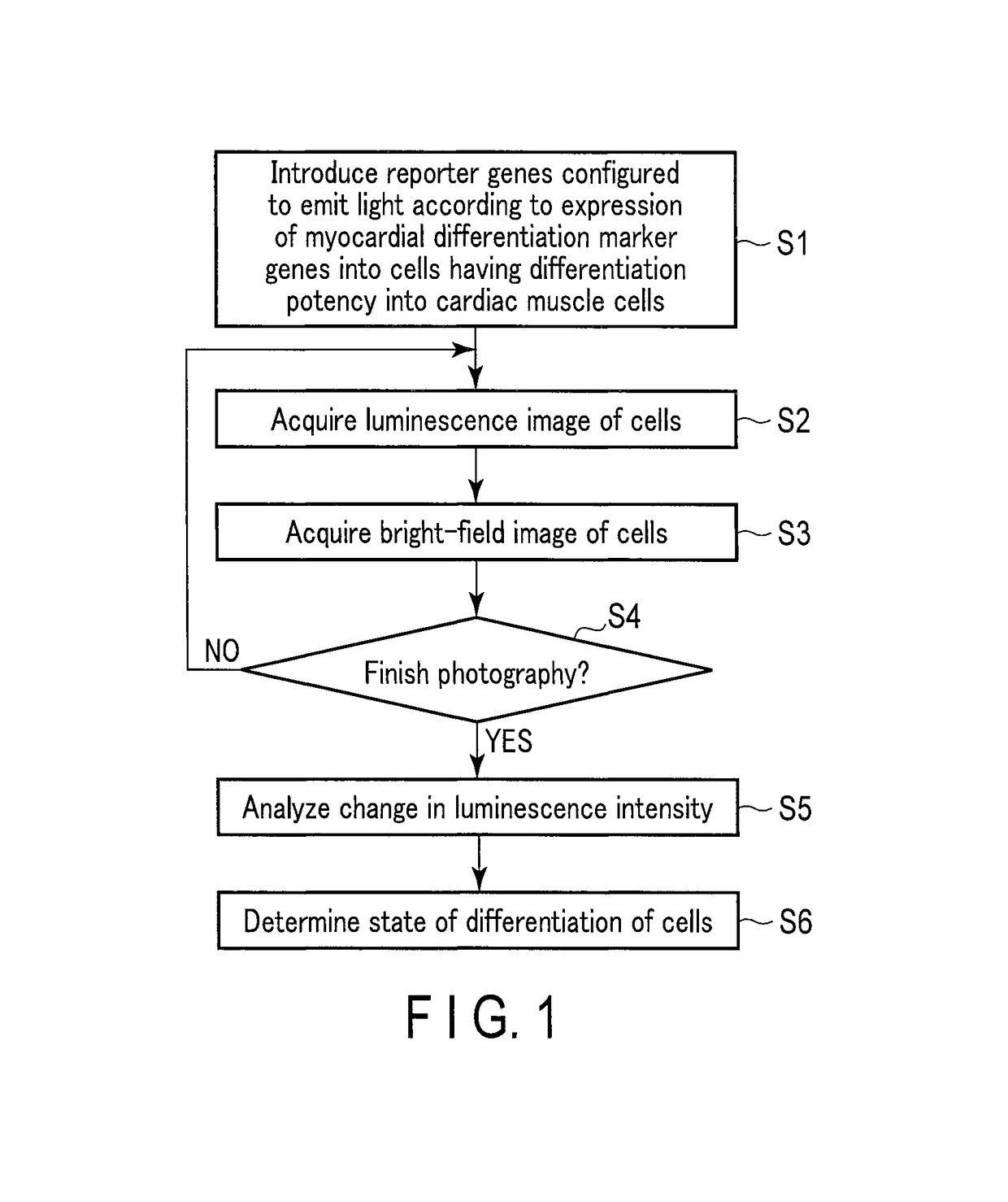
Method for monitoring differentiation into cardiac muscle cells
a technology of cardiac muscle cells and differentiation, applied in the field of monitoring differentiation into cardiac muscle cells, can solve the problems of difficult quantitative observation of gene expression with time for a long period, inability to observe the same target gene expression with time, etc., and achieve the effects of less damage to cells, and reduced amount of luminescent protein included in one cell
- Summary
- Abstract
- Description
- Claims
- Application Information
AI Technical Summary
Benefits of technology
Problems solved by technology
Method used
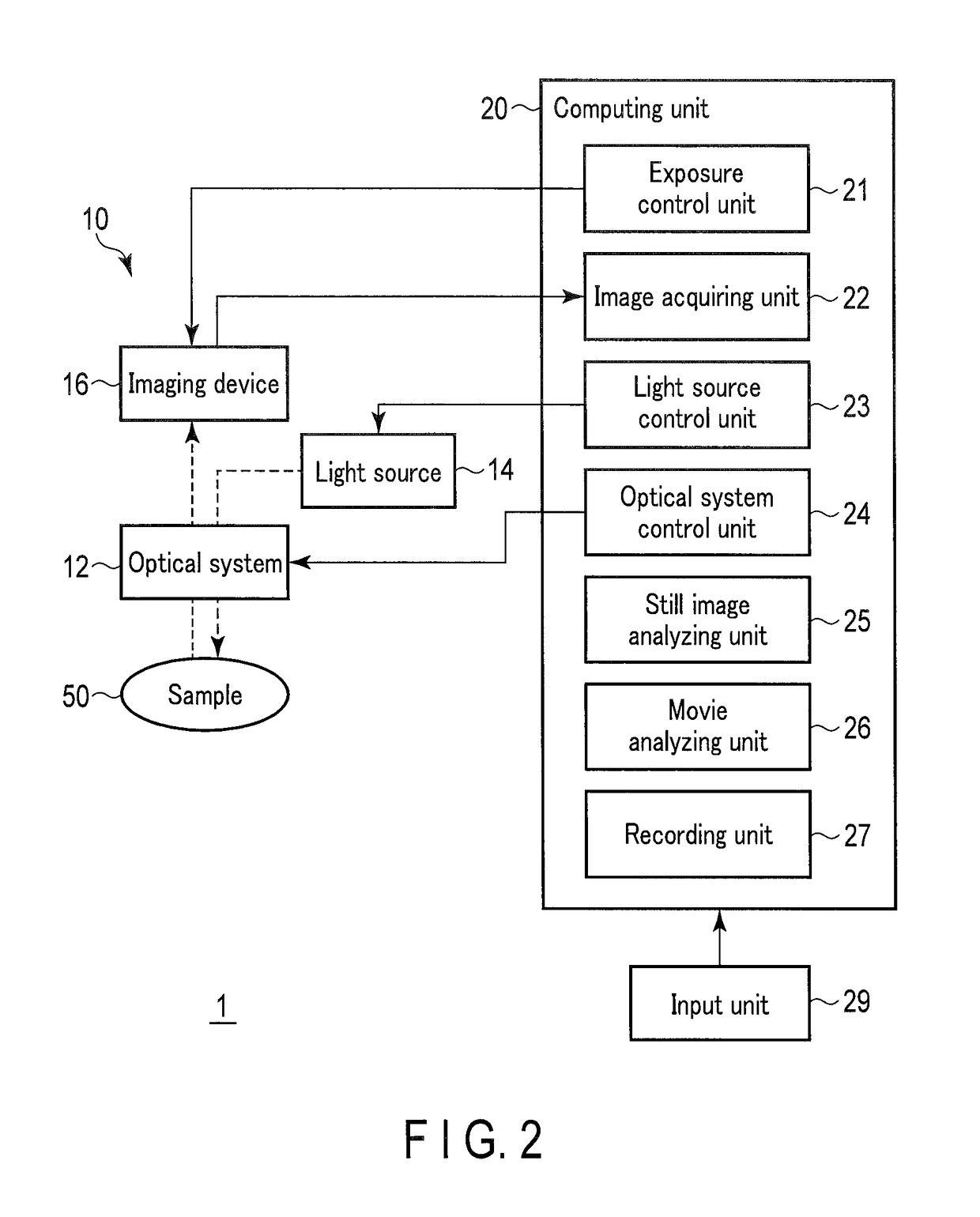
Image

Examples
example 1
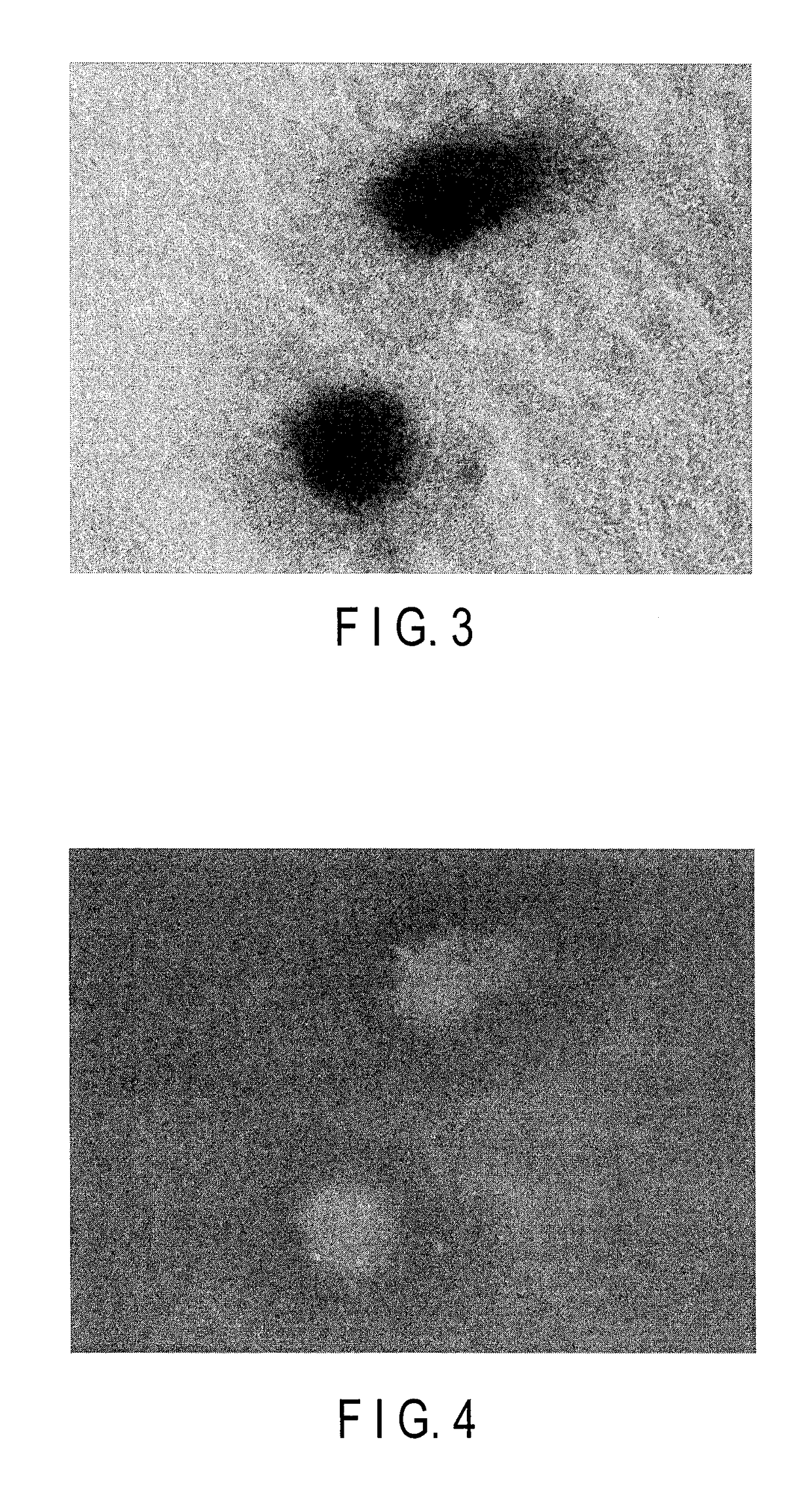
tion of Influence of Autofluorescence in Embryoid Body Derived from Stem Cells
[0127]Mouse ES cells were used to examine the degree of the influence of autofluorescence when excitation light for the excitation of fluorescent protein was applied to an embryoid body derived from stem cells.
[0128](1) Culture of Mouse ES Cells
[0129]For the culture of the mouse ES cells (BRC6, Riken BRC), KO DMEM culture medium (containing 15% of FBS, Penicillin-Streptomycin (SIGMA diluted 100 times), NEAA (SIGMA diluted 100 times), L-Glutamine (final concentration: 2 mM), and 2-mercaptoethanol (final concentration: 0.1 mM)) were used. The mouse ES cells were cultured on a mouse embryonic fibro-blast (MEF cells) whose division was arrested by a mitomycin C treatment.
[0130](2) Formation of Embryoid Body of Mouse ES Cells
[0131]The cultured mouse ES cells were washed with PBS, detached by 0.25% Trypsin-EDTA, and then incubated for 4 hours in an incubator at 37° C. with the KO DMEM culture medium. Feeder cell...
example 2
n Analysis of Cardiac Muscle-Specific Marker in Cardiac Muscle Inducing Process of ES Cells and iPS Cells
[0135]Cardiac troponin T (cTnT) is a protein which expresses specifically in the cardiac muscle. cTnT is utilized as marker genes for myocardial differentiation. The expression of these cTnT genes was used as an index to examine whether the myocardial differentiation inducing process of ES cells and iPS cells can be visualized.
example 2-1
cTnT Expression in Myocardial Differentiation Process of Mouse ES Cells
[0136](1) Production of Mouse ES Cells into which Nucleic Acid Including Promoter Region of cTnT Genes and Luciferase Genes was Introduced
[0137]A promoter region for cTnT genes was cloned with reference to Non Patent Literature “Jin et al., Biochemical and Biophysical Research Communications 1995 Vol. 214, No. 3, p 1168 to 1174”, which is incorporated herein by reference, and the promoter sequence was acquired. In this instance, mouse genomic DNA was used as a template. The following two primers were used.
[0138]
Forward primer 1:(sequence number 1)GCCTCGAGTCTAGACTGAGATACAATGCAAAAGCTGGReverse primer 1:(sequence number 2)GCAGATCTGGTTGAGGGCAGGGCATGGGGAGAGC.
[0139]The acquired promoter sequence for cTnT genes was inserted into a neomycin-resistant pGL4.17 luciferase reporter vector (Promega) to produce a “cTnT gene expression specific luminescent vector pcTnT-GL4”.
[0140]The KO DMEM culture medium was used to culture mo...
PUM
| Property | Measurement | Unit |
|---|---|---|
| time | aaaaa | aaaaa |
| concentration | aaaaa | aaaaa |
| exposure time | aaaaa | aaaaa |
Abstract
Description
Claims
Application Information