
Three-dimensional ultrasound cardiogram four-cavity section image automatic detection method

A slice image and three-dimensional ultrasound technology, applied in ultrasound/sonic/infrasonic image/data processing, organ movement/change detection, image enhancement, etc., can solve problems such as data failure, large amount of calculation for three-dimensional registration, poor real-time performance, etc. , to achieve the effect of large amount of calculation, convenient diagnosis and good real-time performance

Inactive Publication Date: 2007-08-08
SHANGHAI JIAO TONG UNIV
View PDF0 Cites 27 Cited by
  • Summary
  • Abstract
  • Description
  • Claims
  • Application Information

AI Technical Summary

Problems solved by technology

However, all the above-mentioned research work is based on the registration of the three-dimensional cardiogram data of the same person, and can only achieve the registration of two volume data changes within a certain range, and it is often invalid for two volume data with large changes.
3D registration has a large amount of calculation and poor real-time performance

Method used


Image

  • Three-dimensional ultrasound cardiogram four-cavity section image automatic detection method

Examples


Embodiment Construction

[0023] An embodiment of the present invention will be described in detail below in conjunction with the accompanying drawings: this embodiment is implemented on the premise of the technical solution of the present invention, and detailed implementation methods and specific operating procedures are provided, but the protection scope of the present invention is not limited to Examples described below.

[0024] The following is a further detailed description with an example of full volume data collected from any left apex:

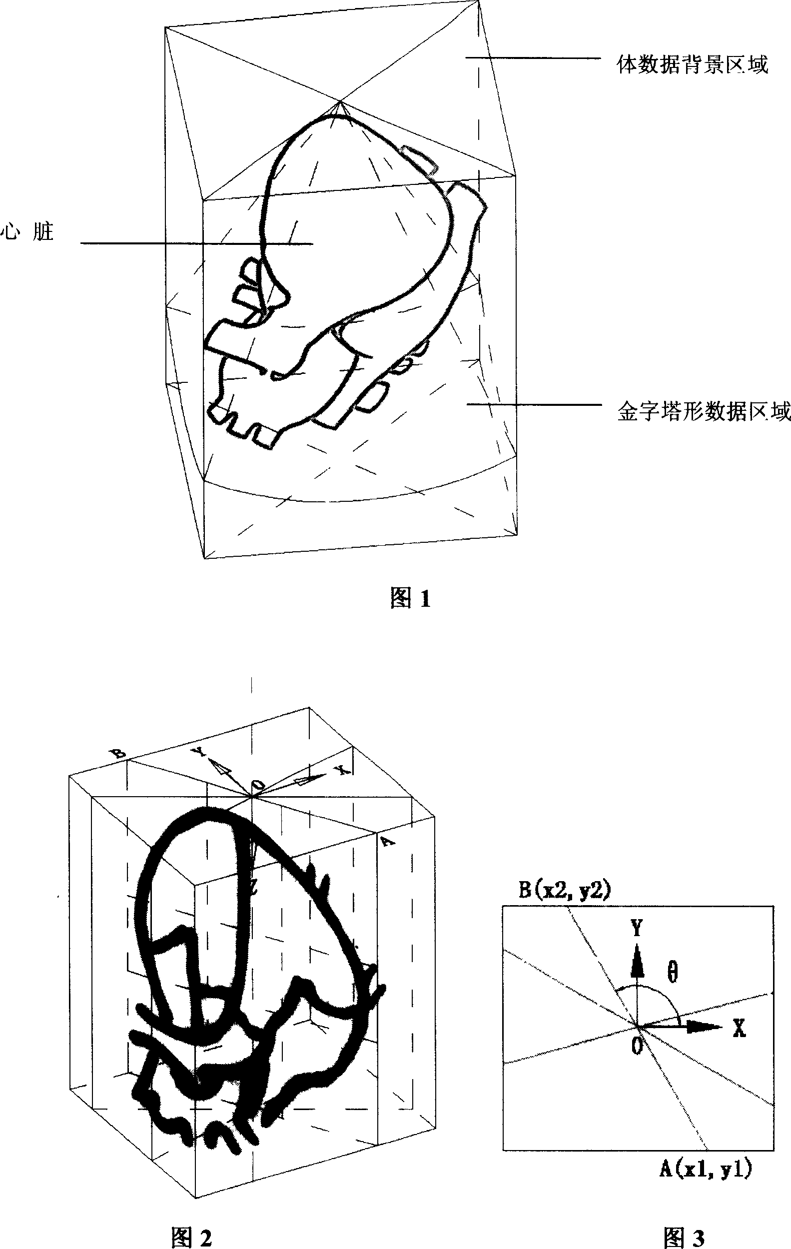
[0025] (1) The three-dimensional matrix (matrix) probe of the Philips Sonos7500 real-time three-dimensional ultrasonic diagnostic instrument is located at the left apex of the heart to collect Full-volume data, and the three-dimensional volume data of the seventh frame at the end of diastole is extracted, and the size of the volume data is 144×160×208 .

[0026] (2) Figure 1 is a schematic diagram of a three-dimensional echocardiogram collected at the left a...


PUM


Abstract

The invention relates to a method for realizing the three-dimension ultrasonic cardiogram four-chamber section image automatic check, which comprises that 1, extracting one three-dimension data from the end-diastolic phase of heart from the heart full-volume data; 2, extracting 180 section images from the three-dimension data to build an image database; 3, calculating out the similarity between each image of database and the template image, to be selected and using wavelet system statistic character to check out the image with highest similarity in the left image, to record the position in the data as the four-chamber watch position. The invention realizes the automatic search function of four-chamber section; therefore, medical staff can use real-time three-dimension ultrasonic technique to diagnose the patient.

Description

technical field [0001] The invention relates to a method in the field of computer application technology, in particular to a method for automatic detection of four-chamber section images of three-dimensional echocardiography. Background technique [0002] Real-time three-dimensional ultrasound system is the main tool for diagnosing heart disease at present. It realizes the function of observing the whole heart structure and movement, and can observe the internal situation of the heart by selecting any slice. At present, doctors mainly use experience in 3D echocardiography to manually find two-dimensional slices one by one to observe the distortion inside the heart. Many literatures have reported the importance of the four-chamber view for the diagnosis of heart disease; for the current clinical diagnosis of real-time three-dimensional echocardiography, doctors also first look for the four-chamber view for observation. However, it is difficult for unskilled doctors to find ...

Claims


Application Information

Patent Timeline
IPC IPC(8): A61B8/13A61B8/08G06T5/00
Inventor 刘小平杨新杨旭朱磊
Owner SHANGHAI JIAO TONG UNIV
Learn More