Method for generating a medical image and corresponding data processing unit and computer software

一种数据处理单元、医学图像的技术,应用在图像数据处理、计算、医药科学等方向,能够解决时间延迟等问题

Active Publication Date: 2008-03-12
SIEMENS HEALTHCARE GMBH
View PDF4 Cites 2 Cited by
  • Summary
  • Abstract
  • Description
  • Claims
  • Application Information

AI Technical Summary

Problems solved by technology

When the user has to sequentially evaluate a large number of medical images produced by means of computed tomography or magnetic resonance tomography (as is the case, for example, with the identification of digital tomographic image data sets), the user may be frustrated by repeated I feel overwhelmed by the work of selecting the appropriate image segment, and this repetitive work will also cause a huge time delay in judging

Method used


Image

  • Method for generating a medical image and corresponding data processing unit and computer software
  • Method for generating a medical image and corresponding data processing unit and computer software
  • Method for generating a medical image and corresponding data processing unit and computer software

Examples


Embodiment Construction

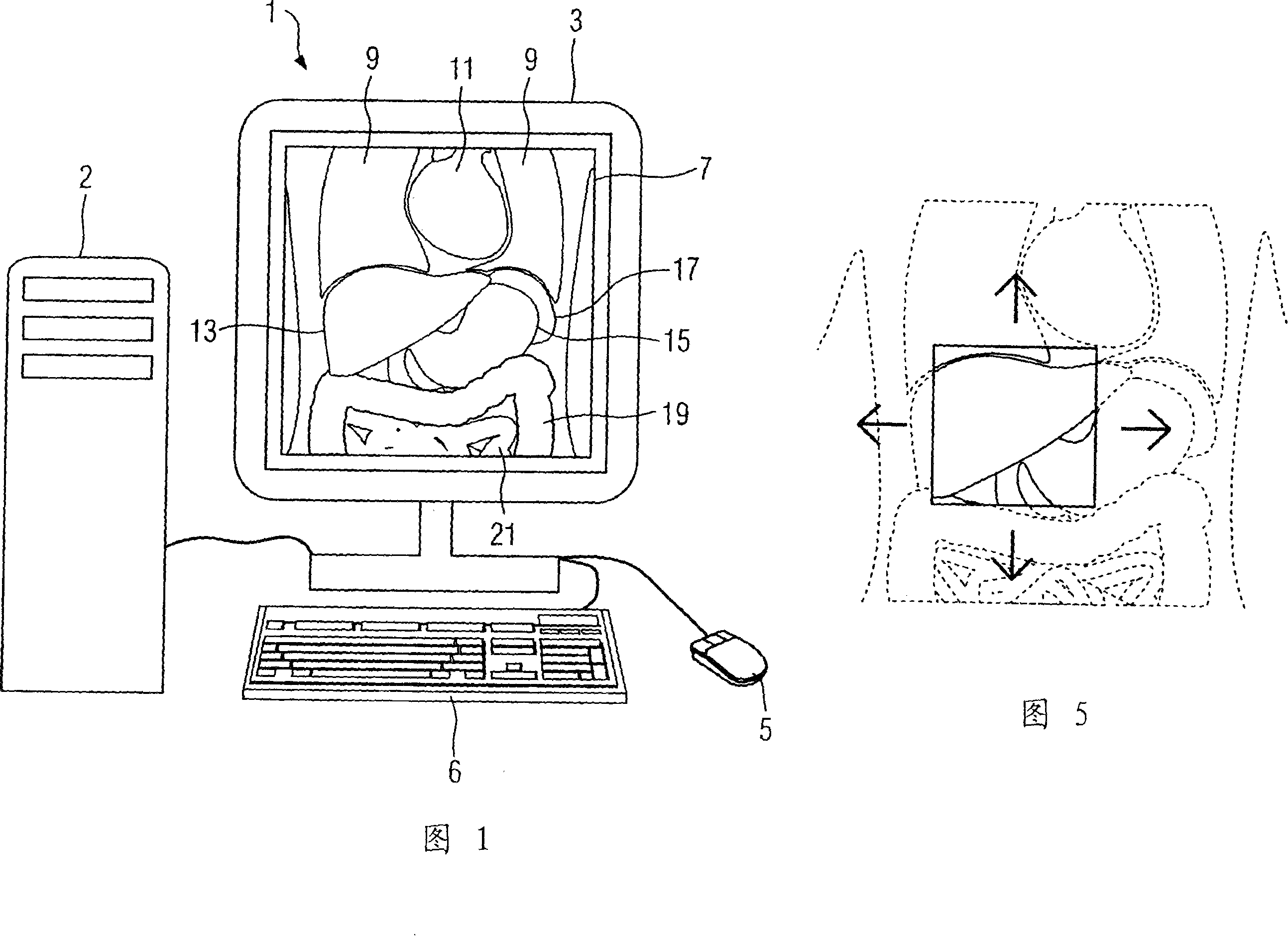
[0031] Fig. 1 shows a display unit 1 suitable for medical images. Such a display unit 1 generally includes a display 3 for displaying medical images for the user and input components such as a keyboard 6 or a mouse 5. By means of these input components, the user can change the display mode of the medical images and follow his own wishes. Adjust it. The display unit 1 is connected to a computer unit 2 which, on the one hand, includes means for managing and connecting to a database, so that recorded data sets and the information contained therein, such as examination instructions, patient data and Imaging means; on the other hand, said computer unit comprises means for processing data sets so that the method of the invention can be implemented in the embodiments described below.

[0032] In order to describe the method of the present invention in detail, FIG. 1 only exemplarily displays the front section 7 of the upper abdomen of the patient to be examined for the user by means...


PUM


Abstract

In a method for generation of a medical image, starting from an acquired data set of an organ to be depicted, a magnification factor and / or an image section to be depicted are automatically determined dependent on the organ to be depicted. Based on this a medical image is generated from the data set. A data processing unit for medical images is fashioned for implementation of such a method and computer software implements such a method when it is executed on a data processing unit.

Description

technical field [0001] The invention relates to a method of generating medical images and a corresponding data processing unit and computer software. Background technique [0002] When judging medical images, users (such as doctors) must browse and judge a large number of images in the shortest possible time. During this process, the user usually prioritizes his attention on the organ that is decisive for the diagnosis of his medical problem. Since important medical information is often hidden in a small area of ​​medical images, and most of the current medical images are displayed in the form of digital images, users will enlarge the corresponding image segments in order to make a detailed analysis of the information. more efficient diagnosis. When the user has to sequentially evaluate a large number of medical images produced by means of computed tomography or magnetic resonance tomography (as is the case, for example, with the identification of digital tomographic image...

Claims


Application Information

Patent Timeline
Patent Type & Authority Applications(China)
IPC IPC(8): A61B6/00A61B19/00G01T1/161G09G5/00
CPCG06T2207/10072G06T7/0012G06T2207/30004
Inventor 斯蒂芬·阿斯曼奥肯·伊金西
Owner SIEMENS HEALTHCARE GMBH
Why Eureka
Try Eureka