
Heart electric function imaging method based on jumping heart model

A technology of functional imaging and cardiac electrophysiology, which is applied in the fields of electrical digital data processing, medical science, special data processing applications, etc. It can solve the problems of inverse problem solution performance degradation, and achieve the effect of overcoming geometric errors.

Inactive Publication Date: 2008-10-15
浙江华际天府智能科技有限公司
View PDF0 Cites 16 Cited by
  • Summary
  • Abstract
  • Description
  • Claims
  • Application Information

AI Technical Summary

Problems solved by technology

When the heart is assumed to be at rest, ignoring the geometric displacement of the heart introduces geometric errors, which will lead to a decrease in the performance of the inverse problem solution

Method used


Image

  • Heart electric function imaging method based on jumping heart model
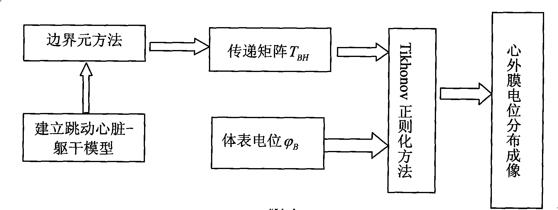
  • Heart electric function imaging method based on jumping heart model
  • Heart electric function imaging method based on jumping heart model

Examples


Embodiment Construction

[0021] A cardiac electrical function imaging method based on a dynamic cardiac model proposed by the present invention is used for imaging epicardial potential distribution information to detect cardiac electrical activity information. The specific implementation steps are as follows:

[0022] (1) The geometric information of the heart, lungs, and torso of the human body is obtained by CT, and the triangular mesh is divided to establish a heart-torso model. The body surface is divided into 412 nodes and 820 meshes; the heart surface is composed of 339 nodes and 654 triangle meshes; the lung is composed of 297 nodes and 586 triangle meshes.

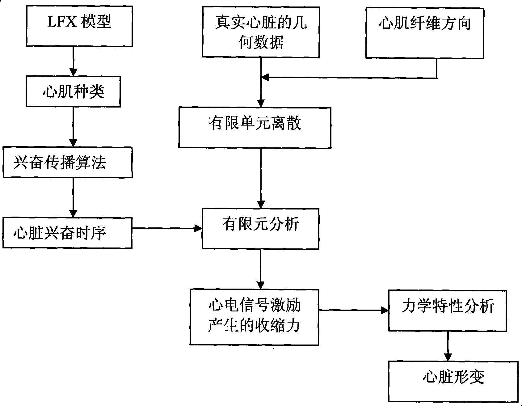
[0023] (2) Using the finite element method, fully considering the fiber rotation of the myocardium, the ventricle is divided into 13 layers of nodes, 1979 units, and 5937 degrees of freedom. Then, according to the type of myocardium in the LFX model and the excitation propagation algorithm, the distribution of the time series of myocardial...


PUM


Abstract

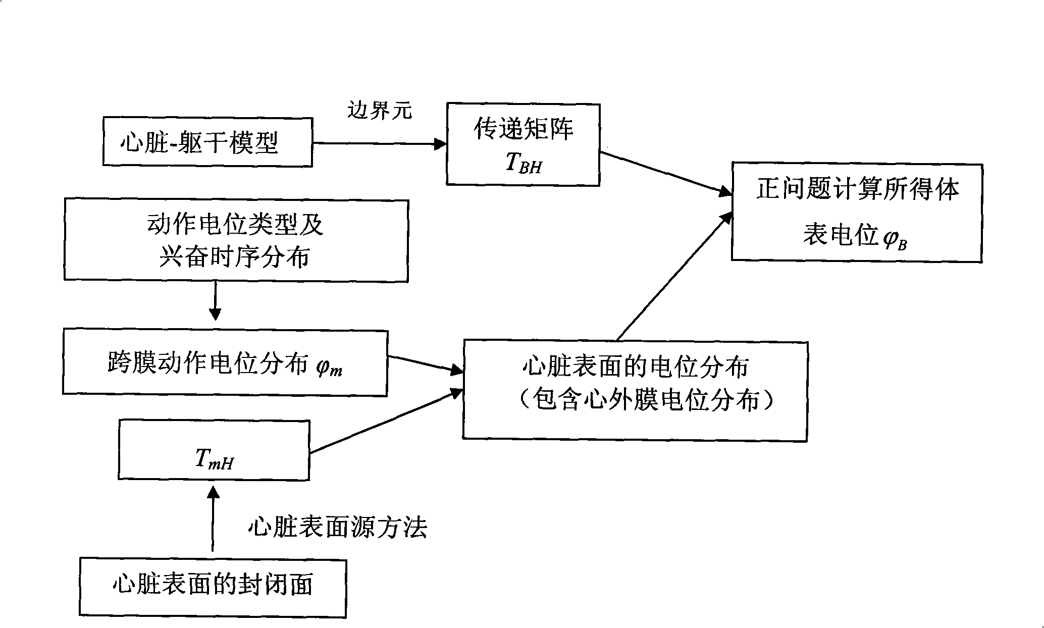
The invention discloses a heart electrical function imaging method based on beating heart model. The method comprises following steps: obtaining the position displacement change of heart based on double-ventricle electricity-coupled heart model; respectively constructing transfer matrixes TBH between epicardium and body surface potential of beating heart at different moments by adopting boundary element method; obtaining the body surface potential distribution PhiB of the body at different moments, calculating the potential distribution of the epicardium, and achieving heart electrical function imaging; and calculating the potential distribution of the epicardium by adopting normal TiKhonov regularization method. The heart electrical function imaging method based on beating heart model can effectively suppress the geometrical error caused by static heart model to obtain more accurate potential distribution imaging of the epicardium and accurately detect the electrical movement information of the heart, thus providing meaningful clinical information for the prevention and the treatment of heart diseases.

Description

technical field [0001] The invention relates to a cardiac electrical function imaging method, in particular to a cardiac electrical functional imaging method based on a beating heart model. Background technique [0002] Cardiovascular disease remains the leading cause of sudden cardiac arrest, killing an estimated 250,000 people each year in Western countries. The most common cause of sudden cardiac arrest is cardiac arrhythmia (such as ventricular fibrillation, ventricular tachycardia), which causes the heart to lose synchronization and lead to sudden death. If defibrillation or CPR (Cardiopulmonaryresuscitation, CPR) is not implemented within minutes after the arrhythmia, death will be inevitable. Some patients did not have any aura and diagnosis of cardiac disease before sudden death. Nonetheless, a recent study demonstrated that 36% of sudden deaths in young athletes had some form of hypertrophic cardiomyopathy. In addition, those with congestive heart failure disease...

Claims


Application Information

Patent Timeline
IPC IPC(8): A61B5/0402G06F19/00
Inventor 夏灵蒋明峰寿国法
Owner 浙江华际天府智能科技有限公司
Learn More