Mixed organization image full-automatic partition method of virtual colonoscope

A technology for organizing images and automatic segmentation, which is used in image analysis, image enhancement, image data processing, etc.

Inactive Publication Date: 2008-10-29
SHAANXI HI TECH MEDICAL INFORMATION
View PDF0 Cites 13 Cited by
  • Summary
  • Abstract
  • Description
  • Claims
  • Application Information

AI Technical Summary

Problems solved by technology

[0011] The purpose of the present invention is to provide a method for fully automatic segmentation of mixed tissue images of virtual colonoscopy, which overcomes the problems caused by traditional threshold segmentation methods, eliminates the influence of local volume effects, and makes the segmentation results more in line with the actual situation

Method used


Image

  • Mixed organization image full-automatic partition method of virtual colonoscope
  • Mixed organization image full-automatic partition method of virtual colonoscope
  • Mixed organization image full-automatic partition method of virtual colonoscope

Examples


Embodiment Construction

[0035] Currently reported virtual colonoscopy requires the same bowel preparation process as traditional optical colonoscopy before colon CT scanning. However, the method of the present invention is based on a special intestinal preparation process. This intestinal preparation method does not require the patient to completely fast the day before the examination, but allows the patient to eat some liquid food and some enhancers to enhance the bowel movement. The imaging effect of CT. In addition, patients feel more comfortable with this type of bowel preparation compared to traditional bowel preparations.

[0036] 1. Transform the multi-layer DICOM data of abdominal CT scan into volume data;

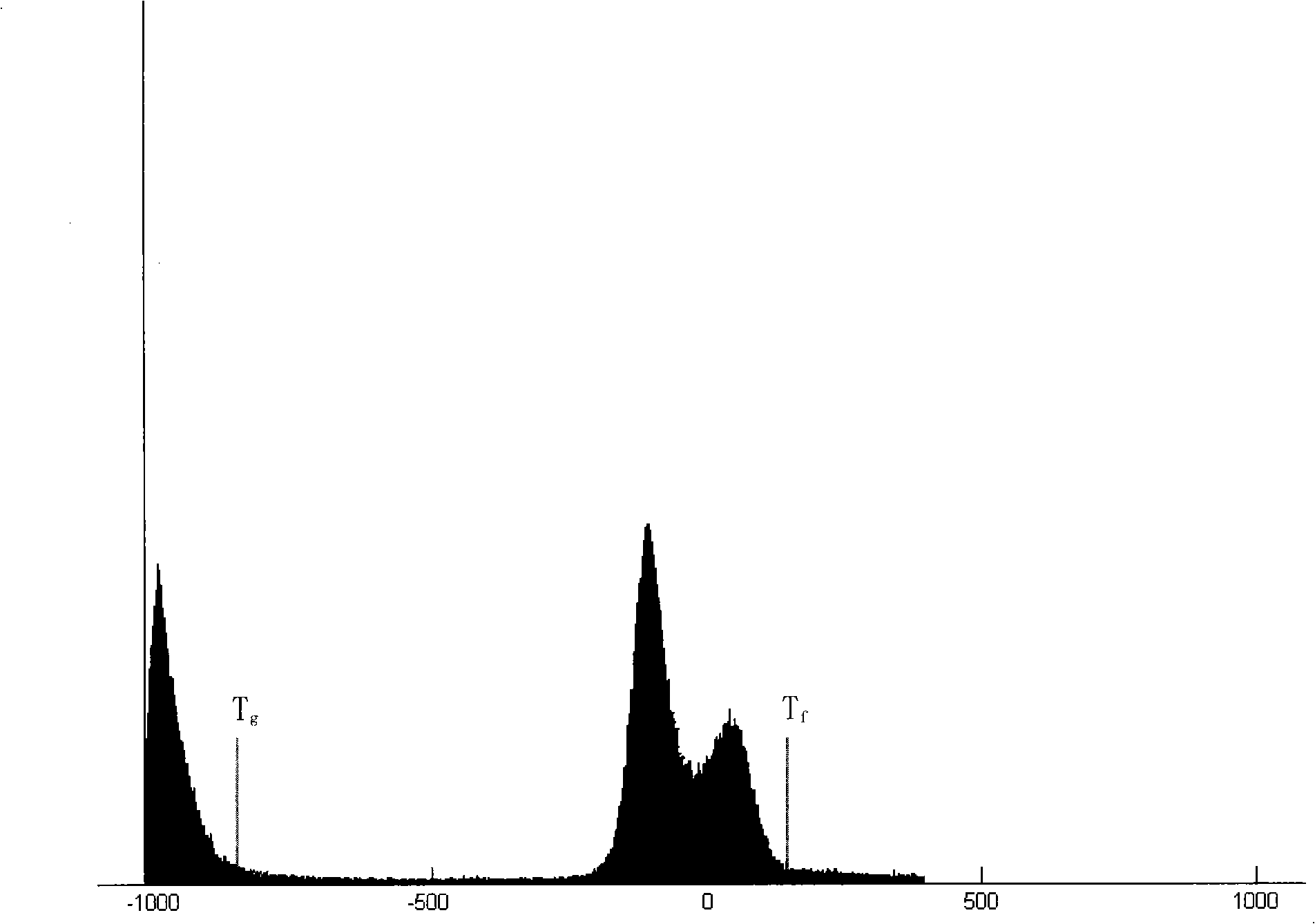
[0037] 2. Calculate the histogram and determine the threshold T according to the histogram g and T f

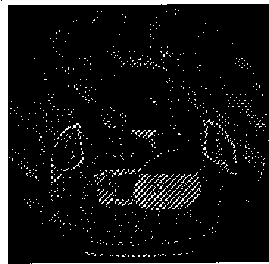
[0038] 3. Perform threshold segmentation;

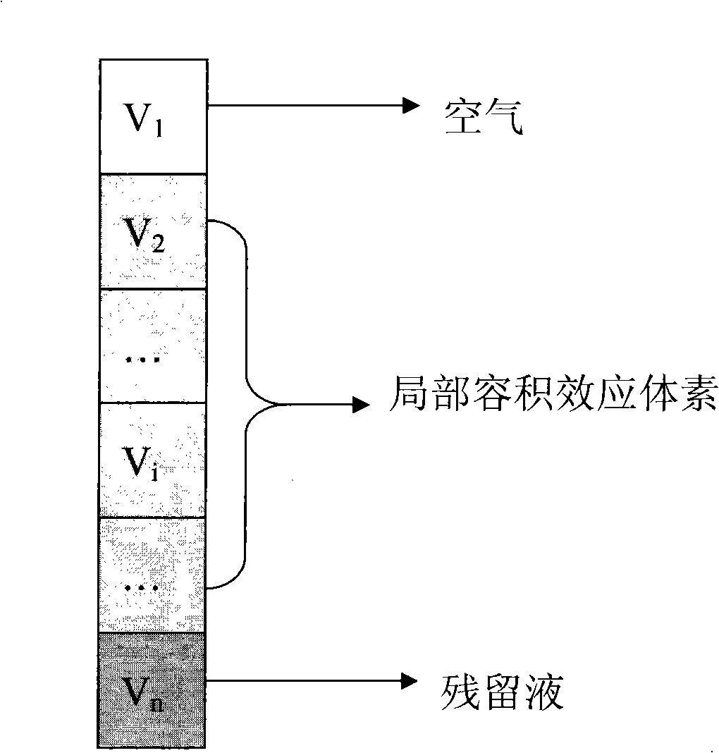
[0039] 4. Use the vertical filter to filter to eliminate the local volume effect voxel between the air and the residual liquid;

[0040]...


PUM


Abstract

The invention provides a full-automatic segmentation method for a mixed tissue image of a simulated colonoscopy, comprising the following steps of: selecting a suitable threshold value of the air and of the enteral residual liquid; carrying out an initial segmentation for the enteral area through a traditional threshold method; wiping a local volume effect between the air and the enteral residual liquid through a vertical filter; conducting an enteral segmentation through a region growing method; enhancing the boundaries of the colon and other issues through a gradient intensity region growing method; repeating a wiping of the local volume effect between the air and the enteral residual liquid through the vertical filter for the inner part of the colon with enhanced boundaries. The full-automatic segmentation method for the mixed tissue image of a simulated colonoscopy overcomes the problems of the traditional threshold segmentation, eliminates the impacts from the local volume effect, and renders the segmentation results better in conformity with the actual conditions.

Description

Technical field: [0001] The invention belongs to the field of medical image processing and computer visualization, in particular to a pre-segmentation method of a virtual colon endoscope. Background technique: [0002] Medical digital endoscopy is also called virtual colonoscopy (Virtual Colonoscopy), which is a new colon disease inspection technology produced with the development of medical imaging equipment and image processing technology. This technique has attracted widespread attention due to its characteristics of simple intestinal preparation, non-invasive and comfortable examination, and broad development prospects. Its principle is to obtain a series of CT (computed tomography) scanning images of the corresponding parts of the patient, and use advanced image processing and visualization technology to directly generate a three-dimensional image inside the lumen from these images for doctors to examine the entire cavity such as polyps, Adenomas and other abnormal mor...

Claims


Application Information

Patent Timeline
Patent Type & Authority Applications(China)
IPC IPC(8): G06T7/00G06T5/00A61B19/00A61B90/00G06T7/136
Inventor 卢虹冰
Owner SHAANXI HI TECH MEDICAL INFORMATION
Why Eureka
Try Eureka